

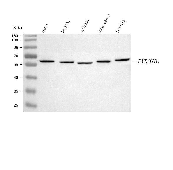
Figure 1. Western blot analysis of PYROXD1 using anti-PYROXD1 antibody (A17214-4). Electrophoresis was performed on a 5-20% SDS-PAGE gel at 70V (Stacking gel) / 90V (Resolving gel) for 2-3 hours. The sample well of each lane was loaded with 30 ug of sample under reducing conditions. Lane 1: human THP-1 whole cell lysates, Lane 2: human SH-SY5Y whole cell lysates, Lane 3: rat brain tissue lysates, Lane 4: mouse brain tissue lysates, Lane 5: mouse NIH/3T3 whole cell lysates. After electrophoresis, proteins were transferred to a nitrocellulose membrane at 150 mA for 50-90 minutes. Blocked the membrane with 5% non-fat milk/TBS for 1.5 hour at RT. The membrane was incubated with rabbit anti-PYROXD1 antigen affinity purified polyclonal antibody (Catalog # A17214-4) at 0.5 microg/mL overnight at 4°C, then washed with TBS-0.1%Tween 3 times with 5 minutes each and probed with a goat anti-rabbit IgG-HRP secondary antibody at a dilution of 1:5000 for 1.5 hour at RT. The signal is developed using an Enhanced Chemiluminescent detection (ECL) kit (Catalog # EK1002) with Tanon 5200 system. A specific band was detected for PYROXD1 at approximately 65 kDa. The expected band size for PYROXD1 is at 50,56,72 kDa.
. PYROXD1 was detected in an immunocytochemical section of U2OS cells. Enzyme antigen retrieval was performed using IHC enzyme antigen retrieval reagent (AR0022) for 15 mins. The cells were blocked with 10% goat serum. And then incubated with 5 microg/mL rabbit anti-PYROXD1 Antibody (A17214-4) overnight at 4°C. DyLight®550 Conjugated Goat Anti-Rabbit IgG (BA1135) was used as secondary antibody at 1:500 dilution and incubated for 30 minutes at 37°C. The section was counterstained with DAPI. Visualize using a fluorescence microscope and filter sets appropriate for the label used.")
. Overlay histogram showing 293T cells stained with A17214-4 (Blue line). To facilitate intracellular staining, cells were fixed with 4% paraformaldehyde and permeabilized with permeabilization buffer. The cells were blocked with 10% normal goat serum. And then incubated with rabbit anti-PYROXD1 Antibody (A17214-4, 1 microg/1x106 cells) for 30 min at 20°C. DyLight®488 conjugated goat anti-rabbit IgG (BA1127, 5-10 microg/1x106 cells) was used as secondary antibody for 30 minutes at 20°C. Isotype control antibody (Green line) was rabbit IgG (1 microg/1x106) used under the same conditions. Unlabelled sample (Red line) was also used as a control.")
Figure 1. Western blot analysis of PYROXD1 using anti-PYROXD1 antibody (A17214-4). Electrophoresis was performed on a 5-20% SDS-PAGE gel at 70V (Stacking gel) / 90V (Resolving gel) for 2-3 hours. The sample well of each lane was loaded with 30 ug of sample under reducing conditions. Lane 1: human THP-1 whole cell lysates, Lane 2: human SH-SY5Y whole cell lysates, Lane 3: rat brain tissue lysates, Lane 4: mouse brain tissue lysates, Lane 5: mouse NIH/3T3 whole cell lysates. After electrophoresis, proteins were transferred to a nitrocellulose membrane at 150 mA for 50-90 minutes. Blocked the membrane with 5% non-fat milk/TBS for 1.5 hour at RT. The membrane was incubated with rabbit anti-PYROXD1 antigen affinity purified polyclonal antibody (Catalog # A17214-4) at 0.5 microg/mL overnight at 4°C, then washed with TBS-0.1%Tween 3 times with 5 minutes each and probed with a goat anti-rabbit IgG-HRP secondary antibody at a dilution of 1:5000 for 1.5 hour at RT. The signal is developed using an Enhanced Chemiluminescent detection (ECL) kit (Catalog # EK1002) with Tanon 5200 system. A specific band was detected for PYROXD1 at approximately 65 kDa. The expected band size for PYROXD1 is at 50,56,72 kDa.
Anti-PYROXD1 Antibody Picoband(r)
A17214-4-BIOTIN
TargetPYROXD1
Product group Antibodies
Overview
- SupplierBoster Bio
- Product NameAnti-PYROXD1 Antibody Picoband(r)
- Delivery Days Customer9
- Concentration500 ug/ml
- Gene ID79912
- Target namePYROXD1
- Target descriptionpyridine nucleotide-disulphide oxidoreductase domain 1
- Target synonymsMFM8; pyridine nucleotide-disulfide oxidoreductase domain-containing protein 1
- Protein IDQ8WU10
- Protein NamePyridine nucleotide-disulfide oxidoreductase domain-containing protein 1
- Scientific DescriptionBoster Bio Anti-PYROXD1 Antibody Picoband® catalog # A17214-4. Tested in ELISA, IF, ICC, WB, Flow Cytometry applications. This antibody reacts with Human, Mouse, Rat. The brand Picoband indicates this is a premium antibody that guarantees superior quality, high affinity, and strong signals with minimal background in Western blot applications. Only our best-performing antibodies are designated as Picoband, ensuring unmatched performance.
- Storage Instruction-20°C,2°C to 8°C
- UNSPSC12352203