


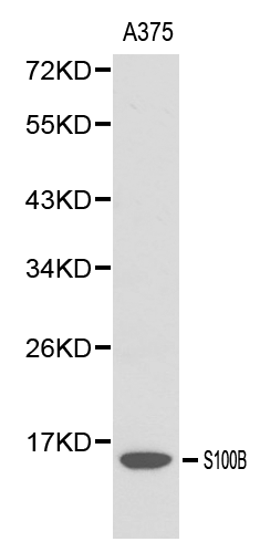
Figure 1. Western blot analysis of S100B using anti-S100B antibody (M00979-1). Electrophoresis was performed on a 5-20% SDS-PAGE gel at 70V (Stacking gel) / 90V (Resolving gel) for 2-3 hours. The sample well of each lane was loaded with 30 ug of sample under reducing conditions. Lane 1: rat brain tissue lysates, Lane 2: rat C6 whole cell lysates, Lane 3: mouse brain tissue lysates, After electrophoresis, proteins were transferred to a nitrocellulose membrane at 150 mA for 50-90 minutes. Blocked the membrane with 5% non-fat milk/TBS for 1.5 hour at RT. The membrane was incubated with rabbit anti-S100B antigen affinity purified monoclonal antibody (Catalog # M00979-1) at 1:500 overnight at 4°C, then washed with TBS-0.1%Tween 3 times with 5 minutes each and probed with a goat anti-rabbit IgG-HRP secondary antibody at a dilution of 1:500 for 1.5 hour at RT. The signal is developed using an Enhanced Chemiluminescent detection (ECL) kit (Catalog # EK1002) with Tanon 5200 system. A specific band was detected for S100B at approximately 11 kDa. The expected band size for S100B is at 11 kDa.


Figure 1. Western blot analysis of S100B using anti-S100B antibody (M00979-1). Electrophoresis was performed on a 5-20% SDS-PAGE gel at 70V (Stacking gel) / 90V (Resolving gel) for 2-3 hours. The sample well of each lane was loaded with 30 ug of sample under reducing conditions. Lane 1: rat brain tissue lysates, Lane 2: rat C6 whole cell lysates, Lane 3: mouse brain tissue lysates, After electrophoresis, proteins were transferred to a nitrocellulose membrane at 150 mA for 50-90 minutes. Blocked the membrane with 5% non-fat milk/TBS for 1.5 hour at RT. The membrane was incubated with rabbit anti-S100B antigen affinity purified monoclonal antibody (Catalog # M00979-1) at 1:500 overnight at 4°C, then washed with TBS-0.1%Tween 3 times with 5 minutes each and probed with a goat anti-rabbit IgG-HRP secondary antibody at a dilution of 1:500 for 1.5 hour at RT. The signal is developed using an Enhanced Chemiluminescent detection (ECL) kit (Catalog # EK1002) with Tanon 5200 system. A specific band was detected for S100B at approximately 11 kDa. The expected band size for S100B is at 11 kDa.
Anti-S100B/S100 Beta Rabbit Monoclonal Antibody
M00979-1
ApplicationsImmunoFluorescence, ImmunoPrecipitation, Western Blot, ImmunoCytoChemistry, ImmunoHistoChemistry
Product group Antibodies
ReactivityHuman, Mouse, Rat
TargetS100B
Overview
- SupplierBoster Bio
- Product NameAnti-S100B/S100 Beta Rabbit Monoclonal Antibody
- Delivery Days Customer9
- ApplicationsImmunoFluorescence, ImmunoPrecipitation, Western Blot, ImmunoCytoChemistry, ImmunoHistoChemistry
- CertificationResearch Use Only
- ClonalityMonoclonal
- Clone IDBCB-19
- Gene ID6285
- Target nameS100B
- Target descriptionS100 calcium binding protein B
- Target synonymsNEF, S100, S100-B, S100beta, protein S100-B, S-100 calcium-binding protein, beta chain, S-100 protein subunit beta, S100 calcium-binding protein, beta (neural)
- HostRabbit
- IsotypeIgG
- Protein IDP04271
- Protein NameProtein S100-B
- Scientific DescriptionBoster Bio Anti-S100B/S100 Beta Rabbit Monoclonal Antibody catalog # M00979-1. Tested in WB, IHC, ICC/IF, IP applications. This antibody reacts with Human, Mouse, Rat.
- ReactivityHuman, Mouse, Rat
- Storage Instruction-20°C
- UNSPSC12352203
References
- Kang WB, Chen YJ, Lu DY, et al. Folic acid contributes to peripheral nerve injury repair by promoting Schwann cell proliferation, migration, and secretion of nerve growth factor. Neural Regen Res. 2019,14(1):132-139. doi: 10.4103/1673-5374.243718Read this paper
- Zhao C, Lin M, Pan Y, et al. Blockage of High-Affinity Choline Transporter Increases Visceral Hypersensitivity in Rats with Chronic Stress. Gastroenterol Res Pract. 2018,2018:9252984. doi: 10.1155/2018/9252984Read this paper
- Ma YH, Zeng X, Qiu XC, et al. Perineurium-like sheath derived from long-term surviving mesenchymal stem cells confers nerve protection to the injured spinal cord. Biomaterials. 2018,160:37-55. doi: 10.1016/j.biomaterials.2018.01.015Read this paper
- Liu H, Lv P, Zhu Y, et al. Salidroside promotes peripheral nerve regeneration based on tissue engineering strategy using Schwann cells and PLGA: in vitro and in vivo. Sci Rep. 2017,7:39869. doi: 10.1038/srep39869Read this paper
- Song XM, Xu XH, Zhu J, et al. Up-regulation of P2X7 receptors mediating proliferation of Schwann cells after sciatic nerve injury. Purinergic Signal. 2015,11(2):203-13. doi: 10.1007/s11302-015-9445-8Read this paper
- Sun T, Ye C, Zhang Z, et al. Cotransplantation of olfactory ensheathing cells and Schwann cells combined with treadmill training promotes functional recovery in rats with contused spinal cords. Cell Transplant. 2013,22 Suppl 1:S27-38. doi: 10.3727/096368913X672118Read this paper
- Lai BQ, Wang JM, Duan JJ, et al. The integration of NSC-derived and host neural networks after rat spinal cord transection. Biomaterials. 2013,34(12):2888-901. doi: 10.1016/j.biomaterials.2012.12.046Read this paper
- Jiang H, Qu W, Han F, et al. Establishment of immortalized Schwann cells derived from rat embryo dorsal root ganglia. Int J Mol Med. 2012,30(3):480-6. doi: 10.3892/ijmm.2012.1016Read this paper
- Wang JM, Zeng YS, Wu JL, et al. Cograft of neural stem cells and schwann cells overexpressing TrkC and neurotrophin-3 respectively after rat spinal cord transection. Biomaterials. 2011,32(30):7454-68. doi: 10.1016/j.biomaterials.2011.06.036Read this paper
- Zhang C, Peng Y, Wang F, et al. A synthetic cantharidin analog for the enhancement of doxorubicin suppression of stem cell-derived aggressive sarcoma. Biomaterials. 2010,31(36):9535-43. doi: 10.1016/j.biomaterials.2010.08.059Read this paper
Datasheet
MSDS
Related products
Product group Antibodies
Anti-S100B AntibodyA28225
ApplicationsWestern Blot, ImmunoHistoChemistry
ReactivityHuman, Mouse, Rat
- SizePrice
Product group Antibodies

Anti-S100B [1C8]Ab02453-1.1
ApplicationsELISA
ReactivityHuman
TargetS100B
- SizePrice
Product group Antibodies
Anti-S100b Antibody130-00023
ApplicationsWestern Blot, ELISA
ReactivityHuman
- SizePrice

Product group Antibodies
Anti-S100B AntibodyAMAB91038
ApplicationsWestern Blot, ImmunoHistoChemistry
ReactivityHuman, Mouse, Rat
TargetS100B
- SizePrice
Product group Antibodies
References
S100B Polyclonal AntibodyBS-2015R
ApplicationsImmunoFluorescence, ELISA, ImmunoCytoChemistry, ImmunoHistoChemistry, ImmunoHistoChemistry Frozen, ImmunoHistoChemistry Paraffin
ReactivityHuman
TargetS100B
- SizePrice
Product group Antibodies

Goat anti-S100BEB12316
ApplicationsWestern Blot, ELISA, ImmunoHistoChemistry
ReactivityHuman
TargetS100B
- SizePrice
Product group Antibodies
S100B AntibodyCSB-PA020643ESR1HU
ApplicationsELISA, ImmunoHistoChemistry
ReactivityHuman
TargetS100B
- SizePrice
Product group Antibodies
ApplicationsImmunoPrecipitation, Western Blot, ImmunoCytoChemistry, ImmunoHistoChemistry
TargetS100B
- SizePrice
Product group Antibodies

ApplicationsWestern Blot, ELISA
ReactivityHuman
TargetS100B
- SizePrice