

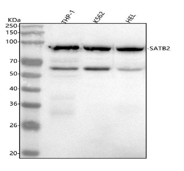
Figure 1. Western blot analysis of SATB2 using anti-SATB2 antibody (M02588). Electrophoresis was performed on a 5-20% SDS-PAGE gel at 70V (Stacking gel) / 90V (Resolving gel) for 2-3 hours. The sample well of each lane was loaded with 30 ug of sample under reducing conditions. Lane 1: human THP-1 whole cell lysates, Lane 2: human K562 whole cell lysates, Lane 3: human HEL whole cell lysates. After electrophoresis, proteins were transferred to a nitrocellulose membrane at 150 mA for 50-90 minutes. Blocked the membrane with 5% non-fat milk/TBS for 1.5 hour at RT. The membrane was incubated with rabbit anti-SATB2 antigen affinity purified monoclonal antibody (Catalog # M02588) at 1:500 overnight at 4°C, then washed with TBS-0.1%Tween 3 times with 5 minutes each and probed with a goat anti-rabbit IgG-HRP secondary antibody at a dilution of 1:1000 for 1.5 hour at RT. The signal is developed using an Enhanced Chemiluminescent detection (ECL) kit (Catalog # EK1002) with Tanon 5200 system. A specific band was detected for SATB2 at approximately 83 kDa. The expected band size for SATB2 is at 83 kDa.


Figure 1. Western blot analysis of SATB2 using anti-SATB2 antibody (M02588). Electrophoresis was performed on a 5-20% SDS-PAGE gel at 70V (Stacking gel) / 90V (Resolving gel) for 2-3 hours. The sample well of each lane was loaded with 30 ug of sample under reducing conditions. Lane 1: human THP-1 whole cell lysates, Lane 2: human K562 whole cell lysates, Lane 3: human HEL whole cell lysates. After electrophoresis, proteins were transferred to a nitrocellulose membrane at 150 mA for 50-90 minutes. Blocked the membrane with 5% non-fat milk/TBS for 1.5 hour at RT. The membrane was incubated with rabbit anti-SATB2 antigen affinity purified monoclonal antibody (Catalog # M02588) at 1:500 overnight at 4°C, then washed with TBS-0.1%Tween 3 times with 5 minutes each and probed with a goat anti-rabbit IgG-HRP secondary antibody at a dilution of 1:1000 for 1.5 hour at RT. The signal is developed using an Enhanced Chemiluminescent detection (ECL) kit (Catalog # EK1002) with Tanon 5200 system. A specific band was detected for SATB2 at approximately 83 kDa. The expected band size for SATB2 is at 83 kDa.
Anti-SATB2 Rabbit Monoclonal Antibody

M02588
ApplicationsImmunoFluorescence, Western Blot, ImmunoCytoChemistry, ImmunoHistoChemistry
Product group Antibodies
ReactivityHuman, Mouse, Rat
TargetSATB2
Overview
- SupplierBoster Bio
- Product NameAnti-SATB2 Rabbit Monoclonal Antibody
- Delivery Days Customer9
- ApplicationsImmunoFluorescence, Western Blot, ImmunoCytoChemistry, ImmunoHistoChemistry
- CertificationResearch Use Only
- ClonalityMonoclonal
- Clone IDADI-19
- FormulationLiquid
- Gene ID23314
- Target nameSATB2
- Target descriptionSATB homeobox 2
- Target synonymsDNA-binding protein SATB2; GLSS; SATB family member 2; special AT-rich sequence-binding protein 2
- HostRabbit
- IsotypeIgG
- Protein IDQ9UPW6
- Protein NameDNA-binding protein SATB2
- Scientific DescriptionBoster Bio Anti-SATB2 Rabbit Monoclonal Antibody catalog # M02588. Tested in WB, IHC, ICC/IF applications. This antibody reacts with Human, Mouse, Rat.
- ReactivityHuman, Mouse, Rat
- Storage Instruction-20°C
- UNSPSC12352203