

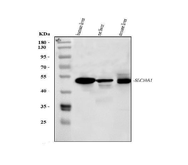
Figure 1. Western blot analysis of SLC10A1 using anti-SLC10A1 antibody (PA1670). Electrophoresis was performed on a 5-20% SDS-PAGE gel at 70V (Stacking gel) / 90V (Resolving gel) for 2-3 hours. The sample well of each lane was loaded with 30 ug of sample under reducing conditions. Lane 1: human liver tissue lysates, Lane 2: rat liver tissue lysates, Lane 3: mouse liver tissue lysates. After electrophoresis, proteins were transferred to a nitrocellulose membrane at 150 mA for 50-90 minutes. Blocked the membrane with 5% non-fat milk/TBS for 1.5 hour at RT. The membrane was incubated with rabbit anti-SLC10A1 antigen affinity purified polyclonal antibody (Catalog # PA1670) at 0.5 microg/mL overnight at 4°C, then washed with TBS-0.1%Tween 3 times with 5 minutes each and probed with a goat anti-rabbit IgG-HRP secondary antibody at a dilution of 1:5000 for 1.5 hour at RT. The signal is developed using an Enhanced Chemiluminescent detection (ECL) kit (Catalog # EK1002) with Tanon 5200 system. A specific band was detected for SLC10A1 at approximately 50 kDa. The expected band size for SLC10A1 is at 38 kDa.
. SLC10A1 was detected in a paraffin-embedded section of rat liver tissue. Heat mediated antigen retrieval was performed in EDTA buffer (pH 8.0, epitope retrieval solution). The tissue section was blocked with 10% goat serum. The tissue section was then incubated with 2 microg/ml rabbit anti-SLC10A1 Antibody (PA1670) overnight at 4°C. Peroxidase Conjugated Goat Anti-rabbit IgG was used as secondary antibody and incubated for 30 minutes at 37°C. The tissue section was developed using HRP Conjugated Rabbit IgG Super Vision Assay Kit (Catalog # SV0002) with DAB as the chromogen.")
. Electrophoresis was performed on a 5-20% SDS-PAGE gel at 70V (Stacking gel) / 90V (Resolving gel) for 2-3 hours. The sample well of each lane was loaded with 30 ug of sample under reducing conditions. Lane 1: rat liver tissue lysates, Lane 2: rat RH35 whole cell lysates, Lane 3: rat kidney tissue lysates, Lane 4: rat brain tissue lysates, Lane 5: mouse liver tissue lysates, Lane 6: mouse HEPA1-6 whole cell lysates, Lane 7: mouse kidney tissue lysates, Lane 8: mouse brain tissue lysates. After electrophoresis, proteins were transferred to a nitrocellulose membrane at 150 mA for 50-90 minutes. Blocked the membrane with 5% non-fat milk/TBS for 1.5 hour at RT. The membrane was incubated with rabbit anti-SLC10A1 antigen affinity purified polyclonal antibody (Catalog # PA1670) at 0.5 microg/mL overnight at 4°C, then washed with TBS-0.1%Tween 3 times with 5 minutes each and probed with a goat anti-rabbit IgG-HRP secondary antibody at a dilution of 1:5000 for 1.5 hour at RT. The signal is developed using an Enhanced Chemiluminescent detection (ECL) kit (Catalog # EK1002) with Tanon 5200 system. A specific band was detected for SLC10A1 at approximately 50 kDa. The expected band size for SLC10A1 is at 38 kDa.")
. SLC10A1 was detected in a paraffin-embedded section of mouse liver tissue. Heat mediated antigen retrieval was performed in EDTA buffer (pH 8.0, epitope retrieval solution). The tissue section was blocked with 10% goat serum. The tissue section was then incubated with 2 microg/ml rabbit anti-SLC10A1 Antibody (PA1670) overnight at 4°C. Peroxidase Conjugated Goat Anti-rabbit IgG was used as secondary antibody and incubated for 30 minutes at 37°C. The tissue section was developed using HRP Conjugated Rabbit IgG Super Vision Assay Kit (Catalog # SV0002) with DAB as the chromogen.")
Figure 1. Western blot analysis of SLC10A1 using anti-SLC10A1 antibody (PA1670). Electrophoresis was performed on a 5-20% SDS-PAGE gel at 70V (Stacking gel) / 90V (Resolving gel) for 2-3 hours. The sample well of each lane was loaded with 30 ug of sample under reducing conditions. Lane 1: human liver tissue lysates, Lane 2: rat liver tissue lysates, Lane 3: mouse liver tissue lysates. After electrophoresis, proteins were transferred to a nitrocellulose membrane at 150 mA for 50-90 minutes. Blocked the membrane with 5% non-fat milk/TBS for 1.5 hour at RT. The membrane was incubated with rabbit anti-SLC10A1 antigen affinity purified polyclonal antibody (Catalog # PA1670) at 0.5 microg/mL overnight at 4°C, then washed with TBS-0.1%Tween 3 times with 5 minutes each and probed with a goat anti-rabbit IgG-HRP secondary antibody at a dilution of 1:5000 for 1.5 hour at RT. The signal is developed using an Enhanced Chemiluminescent detection (ECL) kit (Catalog # EK1002) with Tanon 5200 system. A specific band was detected for SLC10A1 at approximately 50 kDa. The expected band size for SLC10A1 is at 38 kDa.
Anti-Sodium/bile acid cotransporter SLC10A1 Antibody Picoband(r)

PA1670
ApplicationsWestern Blot, ImmunoHistoChemistry
Product group Antibodies
ReactivityChicken, Human, Mouse, Rat
TargetSLC10A1
Overview
- SupplierBoster Bio
- Product NameAnti-SLC10A1 Antibody
- Delivery Days Customer9
- Antibody SpecificityNo cross reactivity with other proteins.
- Application Supplier NoteTested Species: In-house tested species with positive results. Predicted Species: Species predicted to be fit for the product based on sequence similarities. By Heat: Boiling the paraffin sections in 10mM citrate buffer, pH6.0, for 20mins is required for the staining of formalin/paraffin sections. Other applications have not been tested. Optimal dilutions should be determined by end users.
- ApplicationsWestern Blot, ImmunoHistoChemistry
- Applications SupplierIHP, WB, IHC
- CertificationResearch Use Only
- ClonalityPolyclonal
- Concentration500 ug/ml
- FormulationLyophilized
- Gene ID6554
- Target nameSLC10A1
- Target descriptionsolute carrier family 10 member 1
- Target synonymscell growth-inhibiting gene 29 protein; FHCA2; growth-inhibiting protein 29; Na(+)/bile acid cotransporter; Na(+)/taurocholate transport protein; Na/taurocholate cotransporting polypeptide; NTCP; sodium/bile acid cotransporter; sodium/taurocholate cotransporter; sodium/taurocholate cotransporting polypeptide; solute carrier family 10 (sodium/bile acid cotransporter family), member 1; solute carrier family 10 (sodium/bile acid cotransporter), member 1
- HostRabbit
- IsotypeIgG
- Protein IDQ14973
- Protein NameSodium/bile acid cotransporter
- Scientific DescriptionBoster Bio Anti-Sodium/bile acid cotransporter SLC10A1 Antibody catalog # PA1670. Tested in IHC, WB applications. This antibody reacts with Human, Mouse, Rat. The brand Picoband indicates this is a premium antibody that guarantees superior quality, high affinity, and strong signals with minimal background in Western blot applications. Only our best-performing antibodies are designated as Picoband, ensuring unmatched performance.
- ReactivityChicken, Human, Mouse, Rat
- Reactivity SupplierHuman, Mouse, Rat, Chicken
- Storage Instruction-20°C,2°C to 8°C
- UNSPSC12352203
References
- NRF2 and FXR dual signaling pathways cooperatively regulate the effects of oleanolic acid on cholestatic liver injury. Liu J et al., 2023 Jan, PhytomedicineRead more
- Oleanolic acid alleviates ANIT-induced cholestatic liver injury by activating Fxr and Nrf2 pathways to ameliorate disordered bile acids homeostasis. Liu J et al., 2022 Jul 20, PhytomedicineRead more