

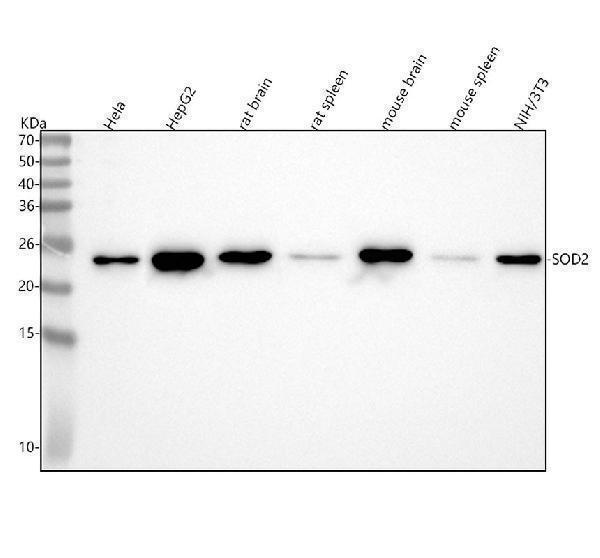
Figure 1. Western blot analysis of SOD2/Mnsod using anti-SOD2/Mnsod antibody (M00349). Electrophoresis was performed on a 5-20% SDS-PAGE gel at 70V (Stacking gel) / 90V (Resolving gel) for 2-3 hours. The sample well of each lane was loaded with 30 ug of sample under reducing conditions. Lane 1: human Hela whole cell lysates, Lane 2: human HepG2 whole cell lysates, Lane 3: rat brain tissue lysates, Lane 4: rat spleen tissue lysates, Lane 5: mouse brain tissue lysates, Lane 6: mouse spleen tissue lysates, Lane 7: mouse NIH/3T3 whole cell lysates. After electrophoresis, proteins were transferred to a nitrocellulose membrane at 150 mA for 50-90 minutes. Blocked the membrane with 5% non-fat milk/TBS for 1.5 hour at RT. The membrane was incubated with rabbit anti-SOD2/Mnsod antigen affinity purified monoclonal antibody (Catalog # M00349) at 1:500 overnight at 4°C, then washed with TBS-0.1%Tween 3 times with 5 minutes each and probed with a goat anti-rabbit IgG-HRP secondary antibody at a dilution of 1:5000 for 1.5 hour at RT. The signal is developed using an Enhanced Chemiluminescent detection (ECL) kit (Catalog # EK1002) with Tanon 5200 system. A specific band was detected for SOD2/Mnsod at approximately 25 kDa. The expected band size for SOD2/Mnsod is at 25 kDa.
. Electrophoresis was performed on a 5-20% SDS-PAGE gel at 70V (Stacking gel) / 90V (Resolving gel) for 2-3 hours. The sample well of each lane was loaded with 30 ug of sample under reducing conditions. Lane 1: rat brain tissue lysates, Lane 2: rat small intestine tissue lysates, Lane 3: mouse brain tissue lysates, Lane 3: mouse small intestine tissue lysates. After electrophoresis, proteins were transferred to a nitrocellulose membrane at 150 mA for 50-90 minutes. Blocked the membrane with 5% non-fat milk/TBS for 1.5 hour at RT. The membrane was incubated with rabbit anti-SOD2/Mnsod antigen affinity purified monoclonal antibody (Catalog # M00349) at 1:500 overnight at 4°C, then washed with TBS-0.1%Tween 3 times with 5 minutes each and probed with a goat anti-rabbit IgG-HRP secondary antibody at a dilution of 1:500 for 1.5 hour at RT. The signal is developed using an Enhanced Chemiluminescent detection (ECL) kit (Catalog # EK1002) with Tanon 5200 system. A specific band was detected for SOD2/Mnsod at approximately 25 kDa. The expected band size for SOD2/Mnsod is at 25 kDa.")
. SOD2 was detected in a paraffin-embedded section of human liver cancer tissue. Heat mediated antigen retrieval was performed in EDTA buffer (pH 8.0, epitope retrieval solution). The tissue section was blocked with 10% goat serum. The tissue section was then incubated with 1:50 rabbit anti-SOD2 Antibody (M00349) overnight at 4°C. Peroxidase Conjugated Goat Anti-rabbit IgG was used as secondary antibody and incubated for 30 minutes at 37°C. The tissue section was developed using HRP Conjugated Rabbit IgG Super Vision Assay Kit (Catalog # SV0002) with DAB as the chromogen.")
. SOD2 was detected in a paraffin-embedded section of human liver cancer tissue. Heat mediated antigen retrieval was performed in EDTA buffer (pH 8.0, epitope retrieval solution). The tissue section was blocked with 10% goat serum. The tissue section was then incubated with 1:50 rabbit anti-SOD2 Antibody (M00349) overnight at 4°C. Peroxidase Conjugated Goat Anti-rabbit IgG was used as secondary antibody and incubated for 30 minutes at 37°C. The tissue section was developed using HRP Conjugated Rabbit IgG Super Vision Assay Kit (Catalog # SV0002) with DAB as the chromogen.")
Figure 1. Western blot analysis of SOD2/Mnsod using anti-SOD2/Mnsod antibody (M00349). Electrophoresis was performed on a 5-20% SDS-PAGE gel at 70V (Stacking gel) / 90V (Resolving gel) for 2-3 hours. The sample well of each lane was loaded with 30 ug of sample under reducing conditions. Lane 1: human Hela whole cell lysates, Lane 2: human HepG2 whole cell lysates, Lane 3: rat brain tissue lysates, Lane 4: rat spleen tissue lysates, Lane 5: mouse brain tissue lysates, Lane 6: mouse spleen tissue lysates, Lane 7: mouse NIH/3T3 whole cell lysates. After electrophoresis, proteins were transferred to a nitrocellulose membrane at 150 mA for 50-90 minutes. Blocked the membrane with 5% non-fat milk/TBS for 1.5 hour at RT. The membrane was incubated with rabbit anti-SOD2/Mnsod antigen affinity purified monoclonal antibody (Catalog # M00349) at 1:500 overnight at 4°C, then washed with TBS-0.1%Tween 3 times with 5 minutes each and probed with a goat anti-rabbit IgG-HRP secondary antibody at a dilution of 1:5000 for 1.5 hour at RT. The signal is developed using an Enhanced Chemiluminescent detection (ECL) kit (Catalog # EK1002) with Tanon 5200 system. A specific band was detected for SOD2/Mnsod at approximately 25 kDa. The expected band size for SOD2/Mnsod is at 25 kDa.
Anti-SOD2/Mnsod Rabbit Monoclonal Antibody

M00349
ApplicationsWestern Blot, ImmunoHistoChemistry
Product group Antibodies
ReactivityHuman, Mouse, Rat
TargetSOD2
Overview
- SupplierBoster Bio
- Product NameAnti-SOD2/Mnsod Rabbit Monoclonal Antibody
- Delivery Days Customer9
- ApplicationsWestern Blot, ImmunoHistoChemistry
- CertificationResearch Use Only
- ClonalityMonoclonal
- Clone IDIDO-19
- FormulationLiquid
- Gene ID6648
- Target nameSOD2
- Target descriptionsuperoxide dismutase 2
- Target synonymsepididymis secretory sperm binding protein; gastric cancer-associated lncRNA 1; GClnc1; indophenoloxidase B; IPOB; IPO-B; manganese-containing superoxide dismutase; mangano-superoxide dismutase; Mn superoxide dismutase; MNSOD; Mn-SOD; MVCD6; superoxide dismutase [Mn], mitochondrial; superoxide dismutase 2, mitochondrial
- HostRabbit
- IsotypeIgG
- Protein IDP04179
- Protein NameSuperoxide dismutase [Mn], mitochondrial
- Scientific DescriptionBoster Bio Anti-SOD2/Mnsod Rabbit Monoclonal Antibody catalog # M00349. Tested in WB, IHC applications. This antibody reacts with Human, Mouse, Rat.
- ReactivityHuman, Mouse, Rat
- Storage Instruction-20°C
- UNSPSC12352203