


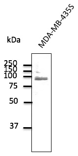
Figure 1. Western blot analysis of TLR2 using anti-TLR2 antibody (M00131). Electrophoresis was performed on a 5-20% SDS-PAGE gel at 70V (Stacking gel) / 90V (Resolving gel) for 2-3 hours. The sample well of each lane was loaded with 30 ug of sample under reducing conditions. Lane 1: human CACO-2 whole cell lysates. After electrophoresis, proteins were transferred to a nitrocellulose membrane at 150 mA for 50-90 minutes. Blocked the membrane with 5% non-fat milk/TBS for 1.5 hour at RT. The membrane was incubated with rabbit anti-TLR2 antigen affinity purified monoclonal antibody (Catalog # M00131) at 1:1000 overnight at 4°C, then washed with TBS-0.1%Tween 3 times with 5 minutes each and probed with a goat anti-rabbit IgG-HRP secondary antibody at a dilution of 1:500 for 1.5 hour at RT. The signal is developed using an Enhanced Chemiluminescent detection (ECL) kit (Catalog # EK1002) with Tanon 5200 system. A specific band was detected for TLR2 at approximately 90 kDa. The expected band size for TLR2 is at 90 kDa.


Figure 1. Western blot analysis of TLR2 using anti-TLR2 antibody (M00131). Electrophoresis was performed on a 5-20% SDS-PAGE gel at 70V (Stacking gel) / 90V (Resolving gel) for 2-3 hours. The sample well of each lane was loaded with 30 ug of sample under reducing conditions. Lane 1: human CACO-2 whole cell lysates. After electrophoresis, proteins were transferred to a nitrocellulose membrane at 150 mA for 50-90 minutes. Blocked the membrane with 5% non-fat milk/TBS for 1.5 hour at RT. The membrane was incubated with rabbit anti-TLR2 antigen affinity purified monoclonal antibody (Catalog # M00131) at 1:1000 overnight at 4°C, then washed with TBS-0.1%Tween 3 times with 5 minutes each and probed with a goat anti-rabbit IgG-HRP secondary antibody at a dilution of 1:500 for 1.5 hour at RT. The signal is developed using an Enhanced Chemiluminescent detection (ECL) kit (Catalog # EK1002) with Tanon 5200 system. A specific band was detected for TLR2 at approximately 90 kDa. The expected band size for TLR2 is at 90 kDa.
Anti-TLR2 Rabbit Monoclonal Antibody
M00131
ApplicationsWestern Blot
Product group Antibodies
ReactivityHuman, Mouse, Rat
TargetTLR2
Overview
- SupplierBoster Bio
- Product NameAnti-TLR2 Rabbit Monoclonal Antibody
- Delivery Days Customer9
- ApplicationsWestern Blot
- CertificationResearch Use Only
- ClonalityMonoclonal
- Clone IDADH-20
- Gene ID7097
- Target nameTLR2
- Target descriptiontoll like receptor 2
- Target synonymsCD282, TIL4, toll-like receptor 2, toll/interleukin-1 receptor-like protein 4
- HostRabbit
- IsotypeIgG
- Protein IDO60603
- Protein NameToll-like receptor 2
- Scientific DescriptionBoster Bio Anti-TLR2 Rabbit Monoclonal Antibody catalog # M00131. Tested in WB application. This antibody reacts with Human, Mouse, Rat.
- ReactivityHuman, Mouse, Rat
- Storage Instruction-20°C
- UNSPSC12352203
References
- Gergen AK, Kohtz PD, Halpern AL, et al. Activation of Toll-Like Receptor 2 Promotes Proliferation of Human Lung Adenocarcinoma Cells. Anticancer Res. 2020,40(10):5361-5369. doi: 10.21873/anticanres.14544Read this paper
- Feng S, Ju L, Shao Z, et al. Therapeutic Effect of C-C Chemokine Receptor Type 1 (CCR1) Antagonist BX471 on Allergic Rhinitis. J Inflamm Res. 2020,13:343-356. doi: 10.2147/JIR.S254717Read this paper
- Xing T, Luo D, Zhao X, et al. Enhanced cytokine expression and upregulation of inflammatory signaling pathways in broiler chickens affected by wooden breast myopathy. J Sci Food Agric. 2021,101(1):279-286. doi: 10.1002/jsfa.10641Read this paper
- Jin X, Yuan Y, Zhang C, et al. Porcine parvovirus nonstructural protein NS1 activates NF-κB and it involves TLR2 signaling pathway. J Vet Sci. 2020,21(3):e50. doi: 10.4142/jvs.2020.21.e50Read this paper
- Peng J, Lin X, Lin H, et al. Up-regulated TLR2 and TLR4 expressions in liver and spleen during acute murine T. gondii infection. Parasitol Res. 2016,115(12):4681-4686.Read this paper
- Wang Y, Ge P, Yang L, et al. Protection of ischemic post conditioning against transient focal ischemia-induced brain damage is associated with inhibition of neuroinflammation via modulation of TLR2 and TLR4 pathways. J Neuroinflammation. 2014,11:15. doi: 10.1186/1742-2094-11-15Read this paper
- Yang L, Xu WG, Xu YP, et al. The effect of peptidoglycan stimulation on basophil-mediated atopic responses during pregnancy and in newborns. J Asthma. 2011,48(4):374-9. doi: 10.3109/02770903.2011.563810Read this paper
Datasheet
MSDS
Related products
Product group Antibodies
Anti-TLR2 AntibodyA121616
ApplicationsWestern Blot
ReactivityCanine, Human, Monkey, Mouse, Rat
- SizePrice
Product group Antibodies
Anti-TLR2 Antibody131-0043
ApplicationsELISA
ReactivityHuman
TargetTLR2
- SizePrice
Product group Antibodies

Anti-TLR2 [TL2.1]Ab01463-10.0
ApplicationsFlow Cytometry, ImmunoFluorescence, ImmunoPrecipitation, Western Blot, Neutralisation/Blocking
ReactivityHuman
TargetTLR2
- SizePrice

Product group Antibodies
Anti-TLR2 AntibodyAMAB91631
ApplicationsWestern Blot
ReactivityHuman
TargetTLR2
- SizePrice
Product group Antibodies
Anti-TLR2 Antibody Picoband(r)A00131-CARRIER-FREE
ApplicationsWestern Blot
ReactivityHuman
TargetTLR2
- SizePrice
Product group Antibodies
References
TLR2 Polyclonal AntibodyBS-1019R
ApplicationsFlow Cytometry, ImmunoFluorescence, Western Blot, ImmunoCytoChemistry, ImmunoHistoChemistry, ImmunoHistoChemistry Frozen, ImmunoHistoChemistry Paraffin
ReactivityHuman, Mouse, Rat
TargetTLR2
- SizePrice
Product group Antibodies
TLR2 AntibodyCSB-PA023601LA01HU
ApplicationsELISA
ReactivityHuman
TargetTLR2
- SizePrice
Product group Antibodies
ApplicationsFlow Cytometry
TargetTLR2
- SizePrice
Product group Antibodies

TLR2 AntibodyLS-C403750
ApplicationsELISA, ImmunoHistoChemistry
ReactivityHuman, Mouse
TargetTLR2
- SizePrice
![TLR2 antibody [N1N2], N-term detects TLR2 protein at cytoplasm on human breast carcinoma by immunohistochemical analysis. Sample: Paraffin-embedded human breast carcinoma. TLR2 antibody [N1N2], N-term (GTX102577) diluted at 1:500.
Antigen Retrieval: Trilogy? (EDTA based, pH 8.0) buffer, 15min](https://www.genetex.com/upload/website/prouct_img/normal/GTX102577/GTX102577_40744_20141128_IHC_w_23060100_281.webp)
Product group Antibodies
TLR2 antibody [N1N2], N-termGTX102577
ApplicationsWestern Blot, ImmunoHistoChemistry, ImmunoHistoChemistry Paraffin
ReactivityHuman
TargetTLR2
- SizePrice