

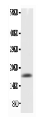
Figure. Western blot analysis of TNF alpha using anti-TNF alpha antibody (RP1000). Electrophoresis was performed on a 5-20% SDS-PAGE gel at 70V(Stacking gel) / 90V (Resolving gel) for 2-3 hours. The sample well of each lane was loaded with 50ug of sample under reducing conditions. Lane: Recombinant Mouse TNFalpha Protein 0.5ng, After Electrophoresis, proteins were transferred to a Nitrocellulose membrane at 150mA for 50-90 minutes. Blocked the membrane with 5% Non-fat Milk/TBS for 1.5 hour at RT. The membrane was incubated with rabbit anti-TNF alpha antigen affinity purified polyclonal antibody (Catalog # RP1000) at 0.5 microg/mL overnight at 4°C, then washed with TBS-0.1%Tween 3 times with 5 minutes each and probed with a goat anti-rabbit IgG-HRP secondary antibody at a dilution of 1:10000 for 1.5 hour at RT. The signal is developed using an Enhanced Chemiluminescent detection (ECL) kit (Catalog # EK1002) with Tanon 5200 system. A specific band was detected for TNF alpha at approximately 17KD. The expected band size for TNF alpha is at 17KD.
Figure. Western blot analysis of TNF alpha using anti-TNF alpha antibody (RP1000). Electrophoresis was performed on a 5-20% SDS-PAGE gel at 70V(Stacking gel) / 90V (Resolving gel) for 2-3 hours. The sample well of each lane was loaded with 50ug of sample under reducing conditions. Lane: Recombinant Mouse TNFalpha Protein 0.5ng, After Electrophoresis, proteins were transferred to a Nitrocellulose membrane at 150mA for 50-90 minutes. Blocked the membrane with 5% Non-fat Milk/TBS for 1.5 hour at RT. The membrane was incubated with rabbit anti-TNF alpha antigen affinity purified polyclonal antibody (Catalog # RP1000) at 0.5 microg/mL overnight at 4°C, then washed with TBS-0.1%Tween 3 times with 5 minutes each and probed with a goat anti-rabbit IgG-HRP secondary antibody at a dilution of 1:10000 for 1.5 hour at RT. The signal is developed using an Enhanced Chemiluminescent detection (ECL) kit (Catalog # EK1002) with Tanon 5200 system. A specific band was detected for TNF alpha at approximately 17KD. The expected band size for TNF alpha is at 17KD.
Anti-TNF alpha Antibody Picoband(r)

RP1000-HRP
ApplicationsWestern Blot, ELISA, ImmunoHistoChemistry
Product group Antibodies
ReactivityMouse
TargetTnf
Overview
- SupplierBoster Bio
- Product NameAnti-TNF alpha Antibody Picoband(r)
- Delivery Days Customer9
- Antibody SpecificityNo cross reactivity with other proteins.
- Application Supplier NoteBy Heat: Boiling the paraffin sections in 10mM citrate buffer, pH6.0, for 20mins is required for the staining of formalin/paraffin sections. Other applications have not been tested. Optimal dilutions should be determined by end users.
- ApplicationsWestern Blot, ELISA, ImmunoHistoChemistry
- CertificationResearch Use Only
- ClonalityPolyclonal
- Concentration500 ug/ml
- ConjugateHRP
- Gene ID21926
- Target nameTnf
- Target descriptiontumor necrosis factor
- Target synonymscachectin; DI; DIF; Tn; TNF-; Tnfa; TNF-a; TNFalpha; TNF-alpha; Tnfs; Tnfsf1a; TNFSF2; Tnlg1f; tumor necrosis factor; tumor necrosis factor ligand 1f; tumor necrosis factor ligand superfamily member 2; tumor necrosis factor-alpha
- HostRabbit
- IsotypeIgG
- Protein IDP06804
- Protein NameTumor necrosis factor
- Scientific DescriptionBoster Bio Anti-TNF alpha Antibody catalog # RP1000. Tested in ELISA, IHC, WB applications. This antibody reacts with Mouse. The brand Picoband indicates this is a premium antibody that guarantees superior quality, high affinity, and strong signals with minimal background in Western blot applications. Only our best-performing antibodies are designated as Picoband, ensuring unmatched performance.
- ReactivityMouse
- Storage Instruction-20°C,2°C to 8°C
- UNSPSC12352203