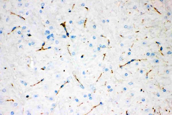
IHC analysis of Tyrosine Hydroxylase using anti-Tyrosine Hydroxylase antibody (MA1100). Tyrosine Hydroxylase was detected in paraffin-embedded section of rat brain tissues. Heat mediated antigen retrieval was performed in citrate buffer (pH6, epitope retrieval solution) for 20 mins. The tissue section was blocked with 10% goat serum. The tissue section was then incubated with 1μg/ml mouse anti-Tyrosine Hydroxylase Antibody (MA1100) overnight at 4°C. Biotinylated goat anti-mouse IgG was used as secondary antibody and incubated for 30 minutes at 37°C. The tissue section was developed using Strepavidin-Biotin-Complex (SABC)(Catalog # SA1021) with DAB as the chromogen.
Anti-Tyrosine Hydroxylase Antibody (Monoclonal, TH-16)

MA1100
Overview
- SupplierBoster Bio
- Product NameAnti-Tyrosine Hydroxylase Antibody (Monoclonal, TH-16)
- Delivery Days Customer9
- Application Supplier NoteOther applications have not been tested. Optimal dilutions should be determined by end users.
- ApplicationsWestern Blot, ImmunoHistoChemistry
- Applications SupplierIHP, IHF, WB, IHC
- CertificationResearch Use Only
- Concentration100 ug/ml
- FormulationLyophilized
- Protein IDP04177
- Protein NameTyrosine 3-monooxygenase
- Scientific DescriptionBoster Bio Anti-Tyrosine Hydroxylase Antibody (Monoclonal, TH-16) catalog # MA1100. Tested in IHC, WB applications. This antibody reacts with Human, Mouse, Rabbit, Rat.
- Storage Instruction-20°C,2°C to 8°C
- UNSPSC12352203
References
- Silencing of Central (Pro)renin Receptor Ameliorates Salt-Induced Renal Injury in Chronic Kidney Disease. Li J et al., 2021 Jul 10, Antioxid Redox SignalRead more

