

anti-VEGF antibody
ARG40735
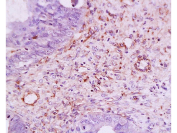
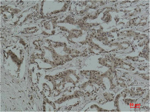
ApplicationsFlow Cytometry, ImmunoFluorescence, ImmunoCytoChemistry, ImmunoHistoChemistry, ImmunoHistoChemistry Paraffin
Product group Antibodies
TargetVEGFA
Overview
- SupplierArigo Biolaboratories
- Product Nameanti-VEGF antibody
- Delivery Days Customer23
- ApplicationsFlow Cytometry, ImmunoFluorescence, ImmunoCytoChemistry, ImmunoHistoChemistry, ImmunoHistoChemistry Paraffin
- CertificationResearch Use Only
- ClonalityPolyclonal
- ConjugateUnconjugated
- Gene ID7422
- Target nameVEGFA
- Target descriptionvascular endothelial growth factor A
- Target synonymsMVCD1; vascular endothelial growth factor A; vascular endothelial growth factor A121; vascular endothelial growth factor A165; vascular permeability factor; VEGF; VPF
- HostRabbit
- IsotypeIgG
- Scientific DescriptionThis gene is a member of the PDGF/VEGF growth factor family and encodes a protein that is often found as a disulfide linked homodimer. This protein is a glycosylated mitogen that specifically acts on endothelial cells and has various effects, including mediating increased vascular permeability, inducing angiogenesis, vasculogenesis and endothelial cell growth, promoting cell migration, and inhibiting apoptosis. Elevated levels of this protein is linked to POEMS syndrome, also known as Crow-Fukase syndrome. Mutations in this gene have been associated with proliferative and nonproliferative diabetic retinopathy. Alternatively spliced transcript variants, encoding either freely secreted or cell-associated isoforms, have been characterized. There is also evidence for the use of non-AUG (CUG) translation initiation sites upstream of, and in-frame with the first AUG, leading to additional isoforms. [provided by RefSeq, Jul 2008]
- Storage Instruction-20°C
- UNSPSC12352203
Related products
Product group Antibodies
References
VEGFA Polyclonal AntibodyBS-0279R
ApplicationsFlow Cytometry, ImmunoFluorescence, Western Blot, ELISA, ImmunoHistoChemistry, ImmunoHistoChemistry Frozen, ImmunoHistoChemistry Paraffin
TargetVEGFA
- SizePrice
Product group Antibodies
ApplicationsNeutralisation/Blocking
TargetVEGFA
- SizePrice
Product group Antibodies

Anti-VEGF [Bevacizumab]Ab00715-10.0
ApplicationsFlow Cytometry, ImmunoPrecipitation, Western Blot, ELISA, Neutralisation/Blocking
TargetVEGFA
- SizePrice
Product group Antibodies
Acetyl Lysine Monoclonal AntibodyCSB-MA080244
ApplicationsWestern Blot, ELISA, ImmunoHistoChemistry
ReactivityAll Species
- SizePrice

Product group Antibodies
Anti-VEGFA AntibodyHPA069116
ApplicationsImmunoHistoChemistry
TargetVEGFA
- SizePrice
Product group Antibodies
Anti-VEGF/VEGFA Antibody Picoband(r)PB9071-CARRIER-FREE
ApplicationsFlow Cytometry, Western Blot, ImmunoHistoChemistry
TargetVEGFA
- SizePrice
Product group Antibodies

Goat anti-VEGFA (aa387-399)EB11645
ApplicationsWestern Blot, ELISA
TargetVEGFA
- SizePrice