

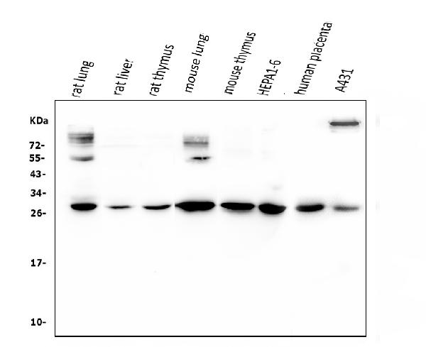
Figure 1. Western blot analysis of VEGF using anti-VEGF antibody (PB9071). Electrophoresis was performed on a 5-20% SDS-PAGE gel at 70V (Stacking gel) / 90V (Resolving gel) for 2-3 hours. The sample well of each lane was loaded with 50ug of sample under reducing conditions. Lane 1: rat lung tissue lysate, Lane 2: rat lver tissue lysate, Lane 3: rat thymus tissue lysate, Lane 4: mouse lung tissue lysate, Lane 5: mouse thymus tissue lysate, Lane 6: HEPA1-6 whole cell lysate, Lane 7: human placenta tissue lysate, Lane 8: A431 whole cell lysate, After Electrophoresis, proteins were transferred to a Nitrocellulose membrane at 150mA for 50-90 minutes. Blocked the membrane with 5% Non-fat Milk/ TBS for 1.5 hour at RT. The membrane was incubated with rabbit anti-VEGF antigen affinity purified polyclonal antibody (Catalog # PB9071) at 0.5 microg/mL overnight at 4°C, then washed with TBS-0.1%Tween 3 times with 5 minutes each and probed with a goat anti-rabbit IgG-HRP secondary antibody at a dilution of 1:10000 for 1.5 hour at RT. The signal is developed using an Enhanced Chemiluminescent detection (ECL) kit (Catalog # EK1002) with Tanon 5200 system. A specific band was detected for VEGF at approximately 27KD. The expected band size for VEGF is at 27KD.
. VEGF was detected in paraffin-embedded section of human rectal cancer tissue. Heat mediated antigen retrieval was performed in EDTA buffer (pH8.0, epitope retrieval solution). The tissue section was blocked with 10% goat serum. The tissue section was then incubated with 1microg/ml rabbit anti-VEGF Antibody (PB9071) overnight at 4°C. Biotinylated goat anti-rabbit IgG was used as secondary antibody and incubated for 30 minutes at 37°C. The tissue section was developed using Strepavidin-Biotin-Complex (SABC)(Catalog # SA1022) with DAB as the chromogen.")
. VEGF was detected in paraffin-embedded section of human rectal cancer tissue. Heat mediated antigen retrieval was performed in EDTA buffer (pH8.0, epitope retrieval solution). The tissue section was blocked with 10% goat serum. The tissue section was then incubated with 1microg/ml rabbit anti-VEGF Antibody (PB9071) overnight at 4°C. Biotinylated goat anti-rabbit IgG was used as secondary antibody and incubated for 30 minutes at 37°C. The tissue section was developed using Strepavidin-Biotin-Complex (SABC)(Catalog # SA1022) with DAB as the chromogen.")
. VEGF was detected in paraffin-embedded section of rat brain tissue. Heat mediated antigen retrieval was performed in EDTA buffer (pH8.0, epitope retrieval solution). The tissue section was blocked with 10% goat serum. The tissue section was then incubated with 1microg/ml rabbit anti-VEGF Antibody (PB9071) overnight at 4°C. Biotinylated goat anti-rabbit IgG was used as secondary antibody and incubated for 30 minutes at 37°C. The tissue section was developed using Strepavidin-Biotin-Complex (SABC)(Catalog # SA1022) with DAB as the chromogen.")
. Overlay histogram showing SiHa cells stained with PB9071 (Blue line). The cells were fixed with 4% paraformaldehyde and blocked with 10% normal goat serum. And then incubated with rabbit anti-VEGF Antibody (PB9071, 1microg/1x106 cells) for 30 min at 20°C. DyLight®488 conjugated goat anti-rabbit IgG (BA1127, 5-10microg/1x106 cells) was used as secondary antibody for 30 minutes at 20°C. Isotype control antibody (Green line) was rabbit IgG (1microg/1x106) used under the same conditions. Unlabelled sample without incubation with primary antibody and secondary antibody (Red line) was used as a blank control.")
Figure 1. Western blot analysis of VEGF using anti-VEGF antibody (PB9071). Electrophoresis was performed on a 5-20% SDS-PAGE gel at 70V (Stacking gel) / 90V (Resolving gel) for 2-3 hours. The sample well of each lane was loaded with 50ug of sample under reducing conditions. Lane 1: rat lung tissue lysate, Lane 2: rat lver tissue lysate, Lane 3: rat thymus tissue lysate, Lane 4: mouse lung tissue lysate, Lane 5: mouse thymus tissue lysate, Lane 6: HEPA1-6 whole cell lysate, Lane 7: human placenta tissue lysate, Lane 8: A431 whole cell lysate, After Electrophoresis, proteins were transferred to a Nitrocellulose membrane at 150mA for 50-90 minutes. Blocked the membrane with 5% Non-fat Milk/ TBS for 1.5 hour at RT. The membrane was incubated with rabbit anti-VEGF antigen affinity purified polyclonal antibody (Catalog # PB9071) at 0.5 microg/mL overnight at 4°C, then washed with TBS-0.1%Tween 3 times with 5 minutes each and probed with a goat anti-rabbit IgG-HRP secondary antibody at a dilution of 1:10000 for 1.5 hour at RT. The signal is developed using an Enhanced Chemiluminescent detection (ECL) kit (Catalog # EK1002) with Tanon 5200 system. A specific band was detected for VEGF at approximately 27KD. The expected band size for VEGF is at 27KD.
Anti-VEGF/VEGFA Antibody Picoband(r)

PB9071
ApplicationsFlow Cytometry, Western Blot, ImmunoHistoChemistry
Product group Antibodies
ReactivityHuman, Mouse, Rat
TargetVEGFA
Overview
- SupplierBoster Bio
- Product NameAnti-VEGF Picoband Antibody
- Delivery Days Customer9
- Antibody SpecificityNo cross reactivity with other proteins.
- Application Supplier NoteWB: The detection limit for VEGF is approximately 0.25ng/lane under reducing conditions. Tested Species: In-house tested species with positive results. Predicted Species: Species predicted to be fit for the product based on sequence similarities. By Heat: Boiling the paraffin sections in 10mM citrate buffer, pH6.0, for 20mins is required for the staining of formalin/paraffin sections. Other applications have not been tested. Optimal dilutions should be determined by end users.
- ApplicationsFlow Cytometry, Western Blot, ImmunoHistoChemistry
- Applications SupplierIHP, WB, IHC
- CertificationResearch Use Only
- ClonalityPolyclonal
- Concentration500 ug/ml
- FormulationLyophilized
- Gene ID7422
- Target nameVEGFA
- Target descriptionvascular endothelial growth factor A
- Target synonymsMVCD1; vascular endothelial growth factor A; vascular endothelial growth factor A121; vascular endothelial growth factor A165; vascular permeability factor; VEGF; VPF
- HostRabbit
- IsotypeIgG
- Protein IDP15692
- Protein NameVascular endothelial growth factor A
- Scientific DescriptionBoster Bio Anti-VEGF/VEGFA Antibody Picoband® catalog # PB9071. Tested in Flow Cytometry, IHC, WB applications. This antibody reacts with Human, Mouse, Rat. The brand Picoband indicates this is a premium antibody that guarantees superior quality, high affinity, and strong signals with minimal background in Western blot applications. Only our best-performing antibodies are designated as Picoband, ensuring unmatched performance.
- ReactivityHuman, Mouse, Rat
- Reactivity SupplierHuman, Rat
- Storage Instruction-20°C,2°C to 8°C
- UNSPSC12352203
References
- MiR-203a-3p inhibits retinal angiogenesis and alleviates proliferative diabetic retinopathy in oxygen-induced retinopathy (OIR) rat model via targeting VEGFA and HIF-1alpha. Han N et al., 2020 Jan, Clin Exp Pharmacol PhysiolRead more
- MGF E peptide improves anterior cruciate ligament repair by inhibiting hypoxia-induced cell apoptosis and accelerating angiogenesis. Sha Y et al., 2019 Jun, J Cell PhysiolRead more