

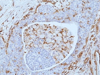
Immunohistochemical staining of formalin-fixed and paraffin embedded human breast cancer tissue sections using anti-VISTA Rabbit Monoclonal Antibody (RM503) at 1:100 dilution.
Immunohistochemical staining of formalin-fixed and paraffin embedded human breast cancer tissue sections using anti-VISTA Rabbit Monoclonal Antibody (RM503) at 1:100 dilution.
anti-VISTA, Rabbit Monoclonal (RM503)
REV-31-1395-00
ApplicationsWestern Blot, ImmunoHistoChemistry
Product group Antibodies
ReactivityHuman, Mouse, Rat
TargetVSIR
Overview
- SupplierRevMAb Biosciences
- Product Nameanti-VISTA, Rabbit Monoclonal (RM503)
- Delivery Days Customer10
- ApplicationsWestern Blot, ImmunoHistoChemistry
- CertificationResearch Use Only
- ClonalityMonoclonal
- Clone IDRM503
- Gene ID64115
- Target nameVSIR
- Target descriptionV-set immunoregulatory receptor
- Target synonymsB7-H5, B7H5, C10orf54, DD1alpha, Dies1, GI24, PD-1H, PP2135, SISP1, VISTA, V-type immunoglobulin domain-containing suppressor of T-cell activation, Death Domain1alpha, PDCD1 homolog, V-domain Ig suppressor of T cell activation, V-set domain-containing immunoregulatory receptor, platelet receptor GI24, sisp-1, stress-induced secreted protein-1
- HostRabbit
- IsotypeIgG
- Protein IDQ9H7M9
- Protein NameV-type immunoglobulin domain-containing suppressor of T-cell activation
- Scientific DescriptionHuman VISTA is a 55 to 65kDa type I Ig membrane protein with the extracellular domain homologous to PD-L1. VISTA is mainly expressed on hematopoietic tissues (spleen, thymus and bone marrow) and on myeloid cells with lower expression on CD4+ and CD8+ T cells. VISTA is a new negative checkpoint regulator that potently suppresses T cell activation. Overexpression of VISTA on tumors inhibits the protective anti-tumor immunity and blockade of VISTA enhances antitumor immunity in multiple tumor models. VISTA on APC delivers negative signaling to T cells via a yet-to-identify binding partner on T cells. - Recombinant Antibody. This antibody reacts to human, mouse or rat VISTA. Isotype: Rabbit IgG. Immunogen: A peptide corresponding to residues near the C-terminus of human VISTA. Application: IHC, WB. Liquid. 50% Glycerol/PBS with 1% BSA and 0.09% sodium azide. Human VISTA is a 55 to 65kDa type I Ig membrane protein with the extracellular domain homologous to PD-L1. VISTA is mainly expressed on hematopoietic tissues (spleen, thymus and bone marrow) and on myeloid cells with lower expression on CD4+ and CD8+ T cells. VISTA is a new negative checkpoint regulator that potently suppresses T cell activation. Overexpression of VISTA on tumors inhibits the protective anti-tumor immunity and blockade of VISTA enhances antitumor immunity in multiple tumor models. VISTA on APC delivers negative signaling to T cells via a yet-to-identify binding partner on T cells.
- ReactivityHuman, Mouse, Rat
- Storage Instruction-20°C,2°C to 8°C
- UNSPSC41116161
Related products
Product group Antibodies

Anti-VISTA [11A4]Ab03306-10.0
ApplicationsWestern Blot, ELISA, ImmunoHistoChemistry
ReactivityHuman
TargetVSIR
- SizePrice
Product group Antibodies
ApplicationsNeutralisation/Blocking
TargetVSIR
- SizePrice
Product group Antibodies
Anti-C10orf54 Antibody144-61872
ApplicationsWestern Blot
ReactivityHuman
TargetVSIR
- SizePrice

Product group Antibodies
Anti-VSIR AntibodyAMAB91253
ApplicationsImmunoHistoChemistry
ReactivityHuman
TargetVSIR
- SizePrice
Product group Antibodies
VISTA Recombinant AntibodyBSM-60327R
ApplicationsImmunoFluorescence, ImmunoHistoChemistry, ImmunoHistoChemistry Frozen, ImmunoHistoChemistry Paraffin
ReactivityHuman
TargetVSIR
- SizePrice
Product group Antibodies
VSIR AntibodyCSB-PA884484LA01HU
ApplicationsWestern Blot, ELISA, ImmunoHistoChemistry
ReactivityHuman, Mouse
TargetVSIR
- SizePrice
Product group Antibodies
Vsir Polyclonal AntibodyCAC09879
ApplicationsWestern Blot, ELISA, ImmunoHistoChemistry
ReactivityMouse
TargetVSIR
- SizePrice
Product group Antibodies
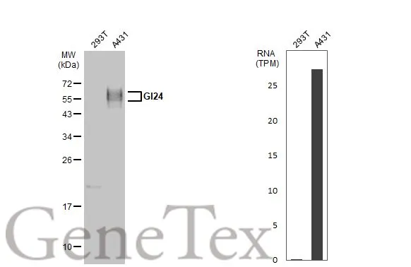
GI24 antibodyGTX104081
ApplicationsWestern Blot
ReactivityHuman
TargetVSIR
- SizePrice
Product group Antibodies

VSIR / GI24 / VISTA AntibodyLS-C783083
ApplicationsWestern Blot
ReactivityHuman
TargetVSIR
- SizePrice