

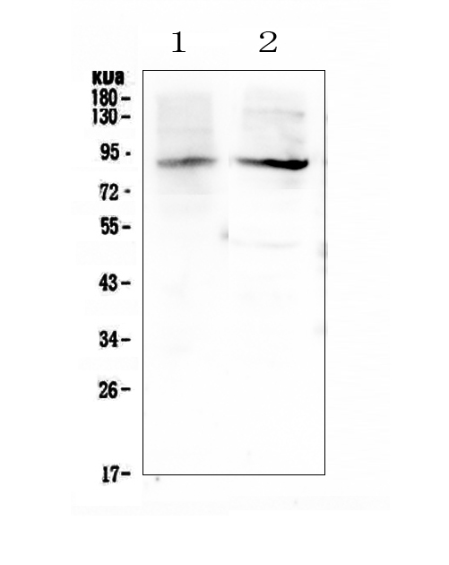
Figure 1. Western blot analysis of XPB using anti-XPB antibody (A03103-1). Electrophoresis was performed on a 5-20% SDS-PAGE gel at 70V (Stacking gel) / 90V (Resolving gel) for 2-3 hours. The sample well of each lane was loaded with 50ug of sample under reducing conditions. Lane 1: rat testis tissue lysates, Lane 2: mouse testis tissue lysates. After Electrophoresis, proteins were transferred to a Nitrocellulose membrane at 150mA for 50-90 minutes. Blocked the membrane with 5% Non-fat Milk/ TBS for 1.5 hour at RT. The membrane was incubated with rabbit anti-XPB antigen affinity purified polyclonal antibody (Catalog # A03103-1) at 0.5 microg/mL overnight at 4°C, then washed with TBS-0.1%Tween 3 times with 5 minutes each and probed with a goat anti-rabbit IgG-HRP secondary antibody at a dilution of 1:10000 for 1.5 hour at RT. The signal is developed using an Enhanced Chemiluminescent detection (ECL) kit (Catalog # EK1002) with Tanon 5200 system. A specific band was detected for XPB at approximately 89KD. The expected band size for XPB is at 89KD.
Figure 1. Western blot analysis of XPB using anti-XPB antibody (A03103-1). Electrophoresis was performed on a 5-20% SDS-PAGE gel at 70V (Stacking gel) / 90V (Resolving gel) for 2-3 hours. The sample well of each lane was loaded with 50ug of sample under reducing conditions. Lane 1: rat testis tissue lysates, Lane 2: mouse testis tissue lysates. After Electrophoresis, proteins were transferred to a Nitrocellulose membrane at 150mA for 50-90 minutes. Blocked the membrane with 5% Non-fat Milk/ TBS for 1.5 hour at RT. The membrane was incubated with rabbit anti-XPB antigen affinity purified polyclonal antibody (Catalog # A03103-1) at 0.5 microg/mL overnight at 4°C, then washed with TBS-0.1%Tween 3 times with 5 minutes each and probed with a goat anti-rabbit IgG-HRP secondary antibody at a dilution of 1:10000 for 1.5 hour at RT. The signal is developed using an Enhanced Chemiluminescent detection (ECL) kit (Catalog # EK1002) with Tanon 5200 system. A specific band was detected for XPB at approximately 89KD. The expected band size for XPB is at 89KD.
Anti-XPB/ERCC3 Antibody Picoband(r)
A03103-1-APC
ApplicationsWestern Blot, ELISA
Product group Antibodies
ReactivityHuman, Mouse, Rat
TargetERCC3
Overview
- SupplierBoster Bio
- Product NameAnti-XPB/ERCC3 Antibody Picoband(r)
- Delivery Days Customer9
- Antibody SpecificityNo cross reactivity with other proteins.
- ApplicationsWestern Blot, ELISA
- CertificationResearch Use Only
- ClonalityPolyclonal
- Concentration500 ug/ml
- ConjugateAPC (Allophycocyanin)
- Gene ID2071
- Target nameERCC3
- Target descriptionERCC excision repair 3, TFIIH core complex helicase subunit
- Target synonymsbasic transcription factor 2 89 kDa subunit; BTF2; BTF2 p89; DNA excision repair protein ERCC-3; DNA repair helicase; DNA repair protein complementing XP-B cells; excision repair cross-complementation group 3; excision repair cross-complementing rodent repair deficiency, complementation group 3; general transcription and DNA repair factor IIH helicase subunit XPB; GTF2H; RAD25; Ssl2; TFIIH; TFIIH 89 kDa subunit; TFIIH basal transcription factor complex 89 kDa subunit; TFIIH basal transcription factor complex helicase XPB subunit; TFIIH p89; TFIIH subunit XPB; TTD2; xeroderma pigmentosum group B-complementing protein; xeroderma pigmentosum, complementation group B; XPB
- HostRabbit
- IsotypeIgG
- Protein IDP19447
- Protein NameGeneral transcription and DNA repair factor IIH helicase subunit XPB
- Scientific DescriptionBoster Bio Anti-XPB/ERCC3 Antibody Picoband® catalog # A03103-1. Tested in ELISA, WB applications. This antibody reacts with Human, Mouse, Rat. The brand Picoband indicates this is a premium antibody that guarantees superior quality, high affinity, and strong signals with minimal background in Western blot applications. Only our best-performing antibodies are designated as Picoband, ensuring unmatched performance.
- ReactivityHuman, Mouse, Rat
- Storage Instruction-20°C,2°C to 8°C
- UNSPSC12352203