

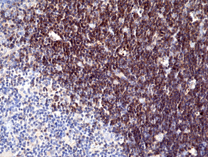
Immunohistochemical staining of formalin fixed and paraffin embedded human thymus tissue section using anti-CD1a rabbit monoclonal antibody (Clone RM393) at a 1:100 dilution.
Immunohistochemical staining of formalin fixed and paraffin embedded human thymus tissue section using anti-CD1a rabbit monoclonal antibody (Clone RM393) at a 1:100 dilution.
anti-CD1a (human), Rabbit Monoclonal (RM393)

REV-31-1279-00
ApplicationsWestern Blot, ImmunoHistoChemistry
Product group Antibodies
ReactivityHuman
TargetCD1A
Overview
- SupplierRevMAb Biosciences
- Product Nameanti-CD1a (human), Rabbit Monoclonal (RM393)
- Delivery Days Customer5
- ApplicationsWestern Blot, ImmunoHistoChemistry
- CertificationResearch Use Only
- ClonalityMonoclonal
- Clone IDRM393
- Gene ID909
- Target nameCD1A
- Target descriptionCD1a molecule
- Target synonymsCD1; CD1A antigen, a polypeptide; cluster of differentiation 1 A; cortical thymocyte antigen CD1A; differentiation antigen CD1-alpha-3; epidermal dendritic cell marker CD1a; FCB6; HTA1; hTa1 thymocyte antigen; R4; T6; T-cell surface antigen T6/Leu-6; T-cell surface glycoprotein CD1a
- HostRabbit
- IsotypeIgG
- Scientific DescriptionCD1a is a non-polymorphic MHC Class 1 related cell surface glycoprotein, expressed in association with Beta 2 microglobulin. CD1a is expressed by cortical thymocytes, Langerhans cells and by interdigitating cells. CD1a is also expressed by some malignancies of T cell lineage and in histiocytosis X. CD1a is expressed on cortical thymocytes, epidermal Langerhans cells, dendritic cells, on certain T-cell leukemias and in various other tissues. CD1a is structurally related to the major histocompatibility complex (MHC) proteins and forms heterodimers with beta-2-microglobulin. The CD1 proteins mediate the presentation of primarily lipid and glycolipid antigens of self or microbial origin to T cells. The CD1 family members are thought to differ in their cellular localization and specificity for particular lipid ligands. CD1a localizes to the plasma membrane and recycles vesicles of the early endocytic system. At least five CD1 genes (CD1a, b, c, d, and e) are identified. - Recombinant Antibody. This antibody reacts to human CD1a. Applications: WB, IHC. Source: Rabbit. Liquid. 50% Glycerol/PBS with 1% BSA and 0.09% sodium azide. CD1a is a non-polymorphic MHC Class 1 related cell surface glycoprotein, expressed in association with Beta 2 microglobulin. CD1a is expressed by cortical thymocytes, Langerhans cells and by interdigitating cells. CD1a is also expressed by some malignancies of T cell lineage and in histiocytosis X. CD1a is expressed on cortical thymocytes, epidermal Langerhans cells, dendritic cells, on certain T-cell leukemias and in various other tissues. CD1a is structurally related to the major histocompatibility complex (MHC) proteins and forms heterodimers with beta-2-microglobulin. The CD1 proteins mediate the presentation of primarily lipid and glycolipid antigens of self or microbial origin to T cells. The CD1 family members are thought to differ in their cellular localization and specificity for particular lipid ligands. CD1a localizes to the plasma membrane and recycles vesicles of the early endocytic system. At least five CD1 genes (CD1a, b, c, d, and e) are identified.
- ReactivityHuman
- Storage Instruction-20°C
- UNSPSC12352203