

Western blot using GeneTexs Affinity Purified anti-AP2A antibody shows detection of a band just below 100 kDa correspond-ing to Human AP2A1 in a various preparations. Lane 1 - HeLa nuclear extract, Lane 2 - HeLa, Lane 3 - 293, Lane 4 - A431 and Lane 5 - Jurkat whole cell lysates. In lanes 6-10 the antibody was preincubated with 1 microg/ml of the immunizing peptide which effect-ively blocks the specific reactivity of this antibody with AP2A. Approximately 20 microg of each lysate was run on a SDS-PAGE and transferred onto nitrocellulose followed by reaction with a 1:500 dilution of anti-AP2A antibody. Detection occurred using a 1:5,000 dilution of HRP-labeled Rabbit anti-Goat IgG for 1 hour at room temperature. A chemi-luminescence system was used for signal detection (Roche) using a 60-sec exposure time.
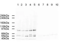
 or without (Lane 1-5) blocking peptide. Lane 1 and 6 : HeLa nuclear extracts Lane 2 and 7 : HeLa whole cell lysates Lane 3 and 8 : 293T whole cell lysates Lane 4 and 9 : A431 whole cell lysates Lane 5 and 10 : Jurkat whole cell lysates Loading : 20 microg Dilution : 1:500")
Western blot using GeneTexs Affinity Purified anti-AP2A antibody shows detection of a band just below 100 kDa correspond-ing to Human AP2A1 in a various preparations. Lane 1 - HeLa nuclear extract, Lane 2 - HeLa, Lane 3 - 293, Lane 4 - A431 and Lane 5 - Jurkat whole cell lysates. In lanes 6-10 the antibody was preincubated with 1 microg/ml of the immunizing peptide which effect-ively blocks the specific reactivity of this antibody with AP2A. Approximately 20 microg of each lysate was run on a SDS-PAGE and transferred onto nitrocellulose followed by reaction with a 1:500 dilution of anti-AP2A antibody. Detection occurred using a 1:5,000 dilution of HRP-labeled Rabbit anti-Goat IgG for 1 hour at room temperature. A chemi-luminescence system was used for signal detection (Roche) using a 60-sec exposure time.
Ap2A antibody
GTX23707
ApplicationsImmunoPrecipitation, Western Blot, ELISA
Product group Antibodies
ReactivityHuman
Overview
- SupplierGeneTex
- Product NameAp2A antibody
- Delivery Days Customer9
- Application Supplier NoteWB: 1:500-1:2000. IP: 1:100. ELISA: 1:5000-1:20000. *Optimal dilutions/concentrations should be determined by the researcher.Not tested in other applications.
- ApplicationsImmunoPrecipitation, Western Blot, ELISA
- CertificationResearch Use Only
- ClonalityPolyclonal
- Concentration1.7 mg/ml
- ConjugateUnconjugated
- HostGoat
- IsotypeIgG
- Scientific DescriptionThe AP2 alpha proteins are found in the AP2 complex in clathrin-coated vesicles. The AP2 complex is a heterotetramer consisting of two large adaptins (alpha or beta), a medium adaptin (mu), and a small adaptin (sigma). The complex is part of the protein coat on the cytoplasmic face of coated vesicles, which link clathrin to receptors in vesicles.
- ReactivityHuman
- Storage Instruction-20°C or -80°C,2°C to 8°C
- UNSPSC12352203