

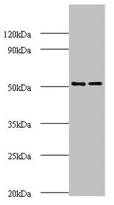
Western blot All lanes: AP-2 complex subunit mu polyclonal Antibody at 2ug/ml Lane 1: Mouse brain tissue Lane 2: Rat brain tissue Secondary Goat polyclonal to rabbit IgG at 1/10000 dilution Predicted band size: 50 kDa Observed band size: 50 kDa


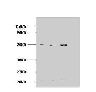
Western blot All lanes: AP-2 complex subunit mu polyclonal Antibody at 2ug/ml Lane 1: Mouse brain tissue Lane 2: Rat brain tissue Secondary Goat polyclonal to rabbit IgG at 1/10000 dilution Predicted band size: 50 kDa Observed band size: 50 kDa
AP2M1 Antibody
CSB-PA00627A0RB
ApplicationsWestern Blot, ELISA, ImmunoHistoChemistry
Product group Antibodies
ReactivityHuman, Mouse, Rat
TargetAP2M1
Overview
- SupplierCusabio
- Product NameAP2M1 Antibody
- Delivery Days Customer20
- ApplicationsWestern Blot, ELISA, ImmunoHistoChemistry
- CertificationResearch Use Only
- ClonalityPolyclonal
- ConjugateUnconjugated
- Gene ID1173
- Target nameAP2M1
- Target descriptionadaptor related protein complex 2 subunit mu 1
- Target synonymsAP50, CLAPM1, MRD60, mu2, AP-2 complex subunit mu, AP-2 mu 2 chain, HA2 50 kDA subunit, adaptin-mu2, adaptor protein complex AP-2 subunit mu, adaptor related protein complex 2 mu 1 subunit, adaptor-related protein complex 2 subunit mu, clathrin adaptor complex AP2, mu subunit, clathrin assembly protein complex 2 medium chain, clathrin assembly protein complex 2 mu medium chain, clathrin coat adaptor protein AP50, clathrin coat assembly protein AP50, clathrin coat-associated protein AP50, clathrin-associated/assembly/adaptor protein, medium 1, plasma membrane adaptor AP-2 50 kDa protein, plasma membrane adaptor AP-2 50kDA protein
- HostRabbit
- IsotypeIgG
- Protein IDQ96CW1
- Protein NameAP-2 complex subunit mu
- Scientific Descriptionomponent of the adaptor protein complex 2 (AP-2). Adaptor protein complexes function in protein transport via transport vesicles in different membrane traffic pathways. Adaptor protein complexes are vesicle coat components and appear to be involved in cargo selection and vesicle formation. AP-2 is involved in clathrin-dependent endocytosis in which cargo proteins are incorporated into vesicles surrounded by clathrin (clathrin-coated vesicles, CCVs) which are destined for fusion with the early endosome. The clathrin lattice serves as a mechanical scaffold but is itself unable to bind directly to membrane components. Clathrin-associated adaptor protein (AP) complexes which can bind directly to both the clathrin lattice and to the lipid and protein components of membranes are considered to be the major clathrin adaptors contributing the CCV formation. AP-2 also serves as a cargo receptor to selectively sort the membrane proteins involved in receptor-mediated endocytosis. AP-2 seems to play a role in the recycling of synaptic vesicle membranes from the presynaptic surface. AP-2 recognizes Y-X-X-[FILMV] (Y-X-X-Phi) and [ED]-X-X-X-L-[LI] endocytosis signal motifs within the cytosolic tails of transmembrane cargo molecules. AP-2 may also play a role in maintaining normal post-endocytic trafficking through the ARF6-regulated, non-clathrin pathway. The AP-2 mu subunit binds to transmembrane cargo proteins; it recognizes the Y-X-X-Phi motifs. The surface region interacting with to the Y-X-X-Phi motif is inaccessible in cytosolic AP-2, but becomes accessible through a conformational change following phosphorylation of AP-2 mu subunit at Tyr-156 in membrane-associated AP-2. The membrane-specific phosphorylation event appears to involve assembled clathrin which activates the AP-2 mu kinase AAK1 By similarity. Plays a role in endocytosis of frizzled family members upon Wnt signaling
- ReactivityHuman, Mouse, Rat
- Storage Instruction-20°C or -80°C
- UNSPSC41116161
Related products
Product group Antibodies
AP2M1 AntibodyCSB-PA00625A0RB
ApplicationsWestern Blot, ELISA, ImmunoHistoChemistry
ReactivityHuman
TargetAP2M1
- SizePrice
Product group Antibodies
Ap2M1 Polyclonal AntibodyCAC07420
ApplicationsWestern Blot, ELISA, ImmunoHistoChemistry
TargetAP2M1
- SizePrice
Product group Antibodies
Anti-AP2M1 Antibody144-02492
ApplicationsWestern Blot, ImmunoHistoChemistry
ReactivityHuman, Mouse, Rat
TargetAP2M1
- SizePrice

Product group Antibodies
Anti-AP2M1 Antibody Picoband(r)A06179-3-CARRIER-FREE
ApplicationsFlow Cytometry, ImmunoFluorescence, Western Blot, ELISA, ImmunoCytoChemistry
ReactivityHuman, Mouse, Rat
TargetAP2M1
- SizePrice
Product group Antibodies
Anti-AP2M1 AntibodyA28295
ApplicationsWestern Blot
ReactivityHuman, Mouse, Rat
- SizePrice
Product group Antibodies

AP50 / AP2M1 AntibodyLS-C748977
ApplicationsImmunoFluorescence
ReactivityHuman
TargetAP2M1
- SizePrice
Product group Antibodies
TargetAP2M1
- SizePrice

Product group Antibodies
Anti-AP2M1 AntibodyHPA036849
ApplicationsImmunoHistoChemistry
ReactivityHuman
TargetAP2M1
- SizePrice
Product group Antibodies
AP2M1 antibodyGTX101502
ApplicationsWestern Blot, ImmunoHistoChemistry, ImmunoHistoChemistry Paraffin
ReactivityHuman
TargetAP2M1
- SizePrice
Product group Antibodies
Anti-AP2M1Y158121
ApplicationsWestern Blot, ELISA, ImmunoHistoChemistry
ReactivityHuman, Mouse, Rat
- SizePrice