

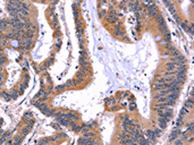
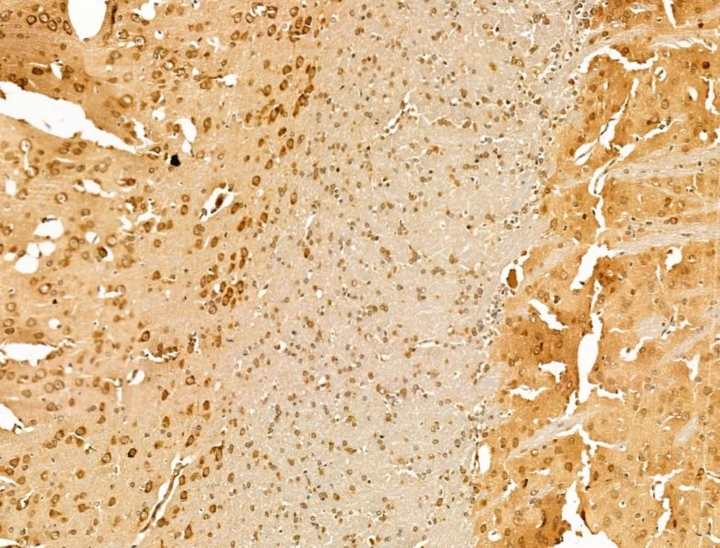
The image on the left is immunohistochemistry of paraffin-embedded Human colon cancer tissue using CSB-PA698018(ARC Antibody) at dilution 1/50, on the right is treated with fusion protein. (Original magnification: x200)
 at dilution 1/50, on the right is treated with fusion protein. (Original magnification: x200)")
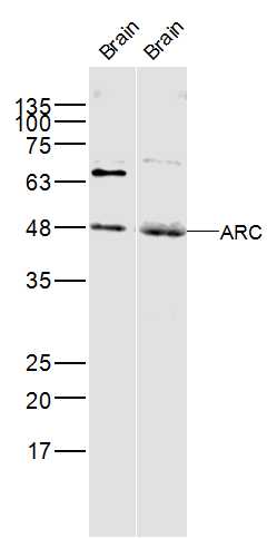
 at dilution 1/597, Secondary antibody: Goat anti rabbit IgG at 1/8000 dilution, Exposure time: 1 minute")
The image on the left is immunohistochemistry of paraffin-embedded Human colon cancer tissue using CSB-PA698018(ARC Antibody) at dilution 1/50, on the right is treated with fusion protein. (Original magnification: x200)
ARC Antibody
CSB-PA698018
ApplicationsWestern Blot, ELISA, ImmunoHistoChemistry
Product group Antibodies
ReactivityHuman, Mouse, Rat
TargetARC
Overview
- SupplierCusabio
- Product NameARC Antibody
- Delivery Days Customer20
- ApplicationsWestern Blot, ELISA, ImmunoHistoChemistry
- CertificationResearch Use Only
- ClonalityPolyclonal
- ConjugateUnconjugated
- Gene ID23237
- Target nameARC
- Target descriptionactivity regulated cytoskeleton associated protein
- Target synonymsArg3.1, hArc, activity-regulated cytoskeleton-associated protein, ARC/ARG3.1, activity-regulated gene 3.1 protein homolog
- HostRabbit
- IsotypeIgG
- Protein IDQ7LC44
- Protein NameActivity-regulated cytoskeleton-associated protein
- Scientific DescriptionArc (for activity-regulated cytoskeleton-associated protein) is a growth factor and immediate early gene that is enriched in brain. Arc mRNA and protein levels are induced by neuronal activity, which is necessary to stimulate neuroplasticity, indicating a potential role for Arc in activity-dependent changes in dendrite function. Arc expression has been detected in neuronal cell bodies and dendrites in the hippocampus, amygdala, hypothalamus, striatum and cortex. Arc has been shown to localize to the cytoskeleton of neuronal cells and appears to colocalize with F-Actin, although it may associate with an Actin-associated protein rather than directly with F-Actin. It has been shown that cocaine-stimulated neuronal activity results in increased Arc mRNA levels in striatum.
- ReactivityHuman, Mouse, Rat
- Storage Instruction-20°C or -80°C
- UNSPSC41116161
Related products

Product group Antibodies
Anti-Arc AntibodyA10637
ApplicationsWestern Blot
ReactivityHuman, Mouse
- SizePrice
Product group Antibodies
Anti-ARC Antibody144-66726
ApplicationsWestern Blot
ReactivityHuman, Mouse
TargetARC
- SizePrice
Product group Antibodies
References
ARC Polyclonal AntibodyBS-0385R
ApplicationsFlow Cytometry, Western Blot, ELISA
ReactivityBovine, Equine, Human, Mouse, Rat
TargetARC
- SizePrice
Product group Antibodies
ApplicationsImmunoPrecipitation, Western Blot, ImmunoCytoChemistry, ImmunoHistoChemistry
TargetARC
- SizePrice
Product group Antibodies

ARC / Arg3.1 AntibodyLS-C401272
ApplicationsELISA, ImmunoHistoChemistry
ReactivityHuman
TargetARC
- SizePrice
Product group Antibodies
ARC antibodyGTX04096
ApplicationsImmunoFluorescence, Western Blot, ImmunoCytoChemistry, ImmunoHistoChemistry, ImmunoHistoChemistry Paraffin
ReactivityHuman, Mouse, Rat
TargetARC
- SizePrice

Product group Antibodies
Anti-ARC AntibodyHPA056430
ApplicationsImmunoCytoChemistry
ReactivityHuman
TargetARC
- SizePrice
Product group Antibodies
Anti-ARCY058421
ApplicationsWestern Blot, ImmunoHistoChemistry
ReactivityHuman, Mouse
- SizePrice
Product group Antibodies
Anti-ARC AntibodyCAB9177
ApplicationsWestern Blot, ELISA
ReactivityHuman
TargetARC
- SizePrice

Product group Antibodies
Anti-Arc Antibody Picoband(r)PB9753-CARRIER-FREE
ApplicationsWestern Blot
ReactivityHuman, Mouse, Rat
TargetARC
- SizePrice