Bio-Connect
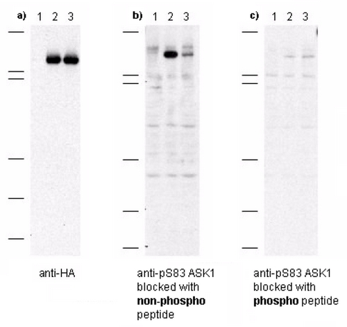
Figure 1. Immunoblot of anti-pS83 ASK1 antibodies shows specificity for phosphorylated human ASK1. Anti-pS83 (aa 76-87) antibody, generated by immunization with phospho peptide coupled to KLH, was tested by immunoblot against lysates of Cos-7 cells after transient transfection, separately, with 1) vector only, 2) recombinant HA-ASK1, and 3) recombinant human HA-ASK1 where S83 was substituted with an alanine residue. Cells were lysed 24 h post-transfection in 200 microL of 1x SDS-sample buffer, heated at 96°C for 5, and vortexed for 30 sec. Samples (10 microL each) were separated on a 12% SDS-PAGE gel and transferred to PVDF (Millipore) followed by blocking for 45 using TTBS supplemented with 5% non-fat dry milk. All incubations were performed at room temperature. In panel a) all samples were incubated with 10 ug/mL mouse anti-HA antibody for 45. After 5X washes with TTBS, reaction with ALP rabbit anti-mouse IgG at 200 ng/mL proceeded for 45 following again by washing as before. The blot was developed using BCIP/NBT. This blot demonstrates both recombinant transfections were successfully over-expressed in the Cos-7 cells. In panel b) all samples were incubated with a 1:1,000 dilution of ASK1 antibody for 45. The antibody was pre-incubated with non-phospho peptide prior to membrane incubation. After 5X washes with TTBS, reaction with HRP goat anti-rabbit IgG at 10 ng/mL proceeded for 45 following again by washing as before. The membrane was processed as before. Lane 2 shows strong specific staining of ASK1. Lane 3, where S83 was replaced with alanine, shows greatly diminished staining. In panel c) all samples were incubated with a 1:1,000 dilution of ASK1 antibody as before except the antibody was preincubated with phospho peptide prior to membrane incubation. No staining is observed after phospho peptide blocking occurs.

ASK1 (MAP3K5) pSer83 Rabbit Polyclonal Antibody

Research Use Only
R1172
OriGene
ApplicationsWestern Blot, ELISA
Product group Antibodies
ReactivityHuman
TargetMAP3K5
200 ug
Sign in to order and to see your custom pricing.
Large volume orders?
Order with a bulk request

Overview

  • Supplier
    OriGene
  • Product Name
    ASK1 (MAP3K5) pSer83 Rabbit Polyclonal Antibody
  • Delivery Days Customer
    14
  • Applications
    Western Blot, ELISA
  • Certification
    Research Use Only
  • Clonality
    Polyclonal
  • Gene ID4217
  • Target name
    MAP3K5
  • Target description
    mitogen-activated protein kinase kinase kinase 5
  • Target synonyms
    apoptosis signal-regulating kinase 1; ASK1; ASK-1; MAP/ERK kinase kinase 5; MAPK/ERK kinase kinase 5; MAPKKK5; MEK kinase 5; MEKK 5; MEKK5; mitogen-activated protein kinase kinase kinase 5
  • Host
    Rabbit
  • Protein IDQ99683
  • Protein Name
    Mitogen-activated protein kinase kinase kinase 5
  • Scientific Description
    ASK1 (MAP3K5) pSer83 rabbit polyclonal antibody, Purified
  • Reactivity
    Human
  • UNSPSC
    12352203