

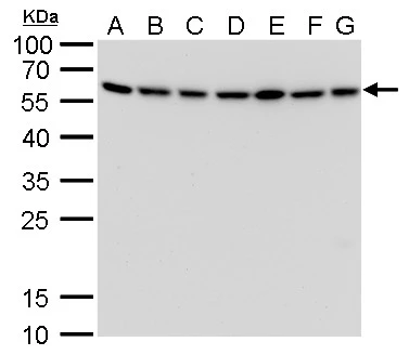
ATG12 antibody [GT166] detects ATG12 protein by western blot analysis. A. 30 μg Neuro2A whole cell lysate/extract B. 30 μg GL261 whole cell lysate/extract C. 30 μg C8D30 whole cell lysate/extract D. 30 μg NIH-3T3 whole cell lysate/extract E. 30 μg BCL-1 whole cell lysate/extract F. 30 μg Raw264.7 whole cell lysate/extract G. 30 μg C2C12 whole cell lysate/extract 12% SDS-PAGE ATG12 antibody [GT166] (GTX629815) dilution: 1:1000 The HRP-conjugated anti-mouse IgG antibody (GTX213111-01) was used to detect the primary antibody.
![ATG12 antibody [GT166] detects ATG12 protein by western blot analysis. A. 30 μg 293T whole cell lysate/extract B. 30 μg A431 whole cell lysate/extract C. 30 μg HeLa whole cell lysate/extract D. 30 μg HepG2 whole cell lysate/extract 12% SDS-PAGE ATG12 antibody [GT166] (GTX629815) dilution: 1:1000 The HRP-conjugated anti-mouse IgG antibody (GTX213111-01) was used to detect the primary antibody.](https://www.genetex.com/upload/website/prouct_img/normal/GTX629815/GTX629815_41463_WB_w_23061202_732.webp "ATG12 antibody [GT166] detects ATG12 protein by western blot analysis. A. 30 μg 293T whole cell lysate/extract B. 30 μg A431 whole cell lysate/extract C. 30 μg HeLa whole cell lysate/extract D. 30 μg HepG2 whole cell lysate/extract 12% SDS-PAGE ATG12 antibody [GT166] (GTX629815) dilution: 1:1000 The HRP-conjugated anti-mouse IgG antibody (GTX213111-01) was used to detect the primary antibody.")
![ATG12 antibody [GT166] detects ATG12 protein at autophagosome by immunofluorescent analysis. Samples: HeLa cells mock (left) and treated with 50μM Chloroquine for 24 hr (right) were fixed in 4% paraformaldehyde at RT for 15 min. Green: ATG12 protein stained by ATG12 antibody [GT166] (GTX629815) diluted at 1:1000. Red: Phalloidin, a F-actin marker.](https://www.genetex.com/upload/website/prouct_img/normal/GTX629815/GTX629815_41463_20141230_IFA_w_23061202_344.webp "ATG12 antibody [GT166] detects ATG12 protein at autophagosome by immunofluorescent analysis. Samples: HeLa cells mock (left) and treated with 50μM Chloroquine for 24 hr (right) were fixed in 4% paraformaldehyde at RT for 15 min. Green: ATG12 protein stained by ATG12 antibody [GT166] (GTX629815) diluted at 1:1000. Red: Phalloidin, a F-actin marker.")
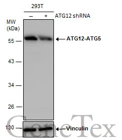
![Non-transfected (–) and transfected (+) 293T whole cell extracts (30 μg) were separated by 10% SDS-PAGE, and the membrane was blotted with ATG12 antibody [GT166] (GTX629815) diluted at 1:500. The HRP-conjugated anti-mouse IgG antibody (GTX213111-01) was used to detect the primary antibody.](https://www.genetex.com/upload/website/prouct_img/normal/GTX629815/GTX629815_41463_20181102_WB_shRNA_watermark_w_23061202_788.webp "Non-transfected (–) and transfected (+) 293T whole cell extracts (30 μg) were separated by 10% SDS-PAGE, and the membrane was blotted with ATG12 antibody [GT166] (GTX629815) diluted at 1:500. The HRP-conjugated anti-mouse IgG antibody (GTX213111-01) was used to detect the primary antibody.")
ATG12 antibody [GT166] detects ATG12 protein by western blot analysis. A. 30 μg Neuro2A whole cell lysate/extract B. 30 μg GL261 whole cell lysate/extract C. 30 μg C8D30 whole cell lysate/extract D. 30 μg NIH-3T3 whole cell lysate/extract E. 30 μg BCL-1 whole cell lysate/extract F. 30 μg Raw264.7 whole cell lysate/extract G. 30 μg C2C12 whole cell lysate/extract 12% SDS-PAGE ATG12 antibody [GT166] (GTX629815) dilution: 1:1000 The HRP-conjugated anti-mouse IgG antibody (GTX213111-01) was used to detect the primary antibody.
ATG12 antibody [GT166]
GTX629815
ApplicationsImmunoFluorescence, Western Blot, ImmunoCytoChemistry, Other Application
Product group Antibodies
ReactivityHuman, Mouse
TargetATG12
Overview
- SupplierGeneTex
- Product NameATG12 antibody [GT166]
- Delivery Days Customer9
- Application Supplier NoteWB: 1:500-1:3000. ICC/IF: 1:100-1:1000. *Optimal dilutions/concentrations should be determined by the researcher.Not tested in other applications.
- ApplicationsImmunoFluorescence, Western Blot, ImmunoCytoChemistry, Other Application
- CertificationResearch Use Only
- ClonalityMonoclonal
- Clone IDGT166
- Concentration1 mg/ml
- ConjugateUnconjugated
- Gene ID9140
- Target nameATG12
- Target descriptionautophagy related 12
- Target synonymsAPG12, APG12L, FBR93, HAPG12, ubiquitin-like protein ATG12, APG12 autophagy 12-like, ATG12 autophagy related 12 homolog, Apg12 (autophagy, yeast) homolog
- HostMouse
- IsotypeIgG2b
- Protein IDO94817
- Protein NameUbiquitin-like protein ATG12
- Scientific DescriptionAutophagy is a process of bulk protein degradation in which cytoplasmic components, including organelles, are enclosed in double-membrane structures called autophagosomes and delivered to lysosomes or vacuoles for degradation. ATG12 is the human homolog of a yeast protein involved in autophagy (Mizushima et al., 1998 [PubMed 9852036]).[supplied by OMIM]
- ReactivityHuman, Mouse
- Storage Instruction-20°C or -80°C,2°C to 8°C
- UNSPSC41116161
Datasheet
Related products
Product group Antibodies
ATG12 AntibodyCSB-PA065886
ApplicationsELISA, ImmunoHistoChemistry
ReactivityHuman
TargetATG12
- SizePrice

Product group Antibodies
Anti-ATG12 Antibody Picoband(r)A00820-2-CARRIER-FREE
ApplicationsFlow Cytometry, Western Blot, ELISA
ReactivityHuman
TargetATG12
- SizePrice
Product group Antibodies
Anti-ATG12 AntibodyA121605
ApplicationsImmunoFluorescence, Western Blot
ReactivityCanine, Human, Monkey, Mouse, Rat
- SizePrice

Product group Antibodies
Anti-ATG12 AntibodyHPA068989
ApplicationsImmunoCytoChemistry
ReactivityHuman
TargetATG12
- SizePrice
Product group Antibodies

APG12 / ATG12 AntibodyLS-C401262
ApplicationsELISA, ImmunoHistoChemistry
ReactivityHuman
TargetATG12
- SizePrice
Product group Antibodies
ATG12 Polyclonal AntibodyBS-4012R
ApplicationsImmunoFluorescence, Western Blot, ELISA, ImmunoCytoChemistry, ImmunoHistoChemistry, ImmunoHistoChemistry Frozen, ImmunoHistoChemistry Paraffin
ReactivityBovine, Canine, Human, Mouse, Porcine, Rabbit, Rat
TargetATG12
- SizePrice
Product group Antibodies
ATG12 antibodyGTX124181
ApplicationsImmunoFluorescence, ImmunoPrecipitation, Western Blot, ImmunoCytoChemistry, ImmunoHistoChemistry, ImmunoHistoChemistry Paraffin
ReactivityHuman, Mouse, Rat
TargetATG12
- SizePrice
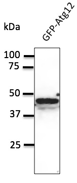
![WB analysis of lysates from (1) Apg12-transfected and (2) non-transfected 293T cells (10 μg per lane) using Apg12 antibody [EPR4800] at a dilution of 1:1,000.](https://www.genetex.com/upload/website/prouct_img/normal/GTX62935/GTX62935_WB_w_23061202_484.webp)
Product group Antibodies
References
ATG12 antibody [EPR4800], C-termGTX62935
ApplicationsImmunoFluorescence, Western Blot, ImmunoCytoChemistry, ImmunoHistoChemistry, ImmunoHistoChemistry Paraffin
ReactivityHuman
TargetATG12
- SizePrice