

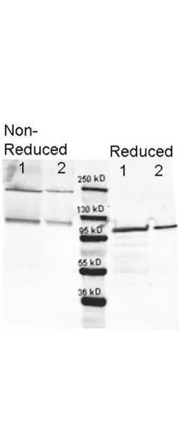
Western blotting using GeneTex anti-beta-Galactosidase antibody (GTX26641). Lane 1 shows 80 ng and lane 2 shows 20 ng loaded onto gel. Results for non-reducing conditions of SDS-PAGE prior to transfer to nitrocellulose are shown on the left side of the figure; results obtainined under reducing conditions are shown on the right. Blots were blocked overnight at 4o C with Blocking Buffer for Fluorescent Western Blotting. The membrane was probed with anti-b-Galactosidase diluted to 1:10,000. Reaction occurred overnight at 4oC. Dylight649? conjugated Gt-a-anti-Rabbit IgG was used for detection. Molecular weight estimation was made by comparison to a prestained MW marker (center).in lane M. Fluorescence image was captured using the VersaDoc Imaging System developed by BIO-RAD. Other detection systems will yield similar results.
 shows detection of a band at ~117 kDa (lane 1) corresponding to b-Gal present in a partially purified preparation (arrowhead). Approximately 1microg of protein was resolved on a 4-20% Tris-Glycine gel by SDS-PAGE and transferred onto nitrocellulose. After blocking, the membrane was probed with the primary antibody diluted to 1:1,000. Reaction occurred overnight at 4o C followed by washes and reaction with a 1:10,000 dilution of IRDyeR 800 conjugated Gt-a-Rabbit IgG (H&L) MX10 for 45 min at room temperature (800 nm channel, green). Molecular weight estimation was made by comparison to prestained MW markers in lane M (700 nm channel, red). IRDye 800 fluorescence image was captured using the Odyssey Infrared Imaging System developed by LI-COR. IRDye is a trademark of LI-COR, Inc. Other detection systems will yield similar results.")
 shows a band at ~117 kDa (lanes 1 - 3) corresponding to 60 ng, 30 ng and 15 ng, respectively of b-Gal present in partially purified preparations (arrowhead). Lane 4 shows no cross reactivity with proteins present in a non-specific control E.coli lysate. Proteins were resolved on a 4-20% Tris-Glycine gel by SDS-PAGE and transferred to nitrocellulose and blocking using Blocking Buffer for Fluorescent Western Blotting. The membrane was probed with fluorescein conjugated anti-b-Galactosidase (GTX26641) diluted to 1:10,000. Reaction occurred for 2 hours at room temperature. Molecular weight estimation was made by comparison to a prestained MW marker in lane M.Fluorescence image was captured using the VersaDoc Imaging System developed by BIO-RAD. Other detection systems will yield similar results.")
. Loading : 1 microg Dilution : 1:1000")
. Lane 1 : 80 ng Lane 2 : 20 ng Dilution : 1:10000")
. Lane 1 : E.coli beta Galactosidase (60 ng) Lane 2 : E.coli beta Galactosidase (30 ng) Lane 3 : E.coli beta Galactosidase (15 ng) Lane 4 : E. coli lysate Dilution : 1:10000")
Western blotting using GeneTex anti-beta-Galactosidase antibody (GTX26641). Lane 1 shows 80 ng and lane 2 shows 20 ng loaded onto gel. Results for non-reducing conditions of SDS-PAGE prior to transfer to nitrocellulose are shown on the left side of the figure; results obtainined under reducing conditions are shown on the right. Blots were blocked overnight at 4o C with Blocking Buffer for Fluorescent Western Blotting. The membrane was probed with anti-b-Galactosidase diluted to 1:10,000. Reaction occurred overnight at 4oC. Dylight649? conjugated Gt-a-anti-Rabbit IgG was used for detection. Molecular weight estimation was made by comparison to a prestained MW marker (center).in lane M. Fluorescence image was captured using the VersaDoc Imaging System developed by BIO-RAD. Other detection systems will yield similar results.
beta Galactosidase antibody (FITC)
GTX26641
ApplicationsImmunoFluorescence, Western Blot, ImmunoCytoChemistry
Product group Antibodies
ReactivityBacteria
Overview
- SupplierGeneTex
- Product Namebeta Galactosidase antibody (FITC)
- Delivery Days Customer9
- Application Supplier NoteWB: 1:10000. ICC/IF: 1:500-1:2500. *Optimal dilutions/concentrations should be determined by the researcher.Not tested in other applications.
- ApplicationsImmunoFluorescence, Western Blot, ImmunoCytoChemistry
- CertificationResearch Use Only
- ClonalityPolyclonal
- Concentration5 mg/ml
- ConjugateFITC
- HostRabbit
- IsotypeIgG
- Scientific DescriptionRepressed during biofilm formation. [More information is available at EcoGene: EG10527]. galacotsidase is a metalloenzyme exhibiting broad substrate specificity. [More information is available at EcoCyc: EG10527].
- ReactivityBacteria
- Storage Instruction-20°C,2°C to 8°C
- UNSPSC12352203