

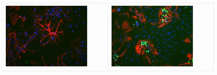
ICC/IF analysis of rat brain neural cultured cells using GTX60996 c-Fos antibody [2H2]. Left : Untreated cells Right : Cells stimulated with membrane deplorization buffer for 5 hours. Green : Primary antibody Red : GFAP antibody Blue : DAPI
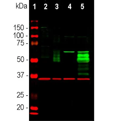
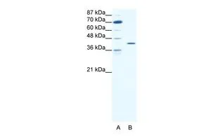
![WB analysis of various samples using GTX60996 c-Fos antibody [2H2]. Lane 1: Protein marker (red) Lane 2: HeLa cells in serum free media Lane 3: HeLa cells stimulated with 20% FBS for 2hrs after 36hrs in serum free media. Lane 4: Rat cortical neurons Lane 5: Rat cortical neurons treated with membrane depolarization buffer for 5hrs. Dilution : 1:1000](https://www.genetex.com/upload/website/prouct_img/normal/GTX60996/GTX60996_20231130_WB_23112922_835.webp "WB analysis of various samples using GTX60996 c-Fos antibody [2H2]. Lane 1: Protein marker (red) Lane 2: HeLa cells in serum free media Lane 3: HeLa cells stimulated with 20% FBS for 2hrs after 36hrs in serum free media. Lane 4: Rat cortical neurons Lane 5: Rat cortical neurons treated with membrane depolarization buffer for 5hrs. Dilution : 1:1000")
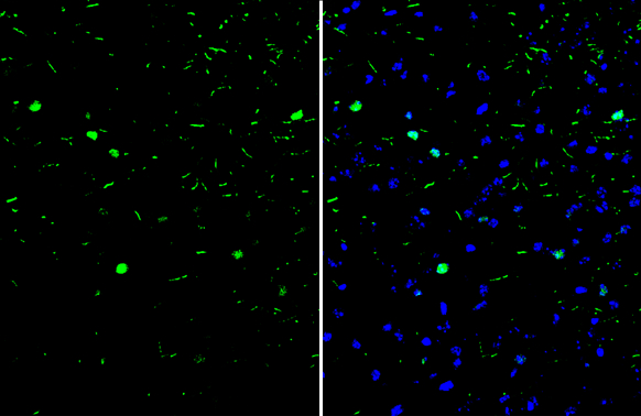
![ICC/IF analysis of HeLa cells using GTX60996 c-Fos antibody [2H2]. Left : Serum-starved HeLa cells Right : Serum-starved and then stimulated with 20% FBS for 2 hours Green : Primary antibody Red : Vimentin antibody Blue : DAPI Dilution : 1:1000](https://www.genetex.com/upload/website/prouct_img/normal/GTX60996/GTX60996_20231130_ICCIF_23112922_697.webp "ICC/IF analysis of HeLa cells using GTX60996 c-Fos antibody [2H2]. Left : Serum-starved HeLa cells Right : Serum-starved and then stimulated with 20% FBS for 2 hours Green : Primary antibody Red : Vimentin antibody Blue : DAPI Dilution : 1:1000")
![IHC-P analysis of rat hippocampus tissue using using GTX60996 c-Fos antibody [2H2] Antigen retrieval : Citrate buffer Dilution : 1:1000](https://www.genetex.com/upload/website/prouct_img/normal/GTX60996/GTX60996_20231130_IHC-P_23112922_526.webp "IHC-P analysis of rat hippocampus tissue using using GTX60996 c-Fos antibody [2H2] Antigen retrieval : Citrate buffer Dilution : 1:1000")
![IHC-P analysis of mouse hippocampus tissue using using GTX60996 c-Fos antibody [2H2] Antigen retrieval : Citrate buffer Dilution : 1:1000](https://www.genetex.com/upload/website/prouct_img/normal/GTX60996/GTX60996_20231130_IHC-P_1_23112922_783.webp "IHC-P analysis of mouse hippocampus tissue using using GTX60996 c-Fos antibody [2H2] Antigen retrieval : Citrate buffer Dilution : 1:1000")
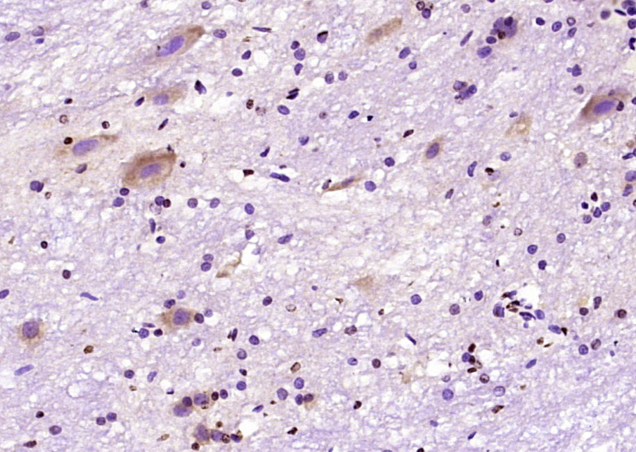
![c-Fos antibody [2H2] detects c-Fos protein by immunohistochemical analysis. Sample: Paraffin-embedded mouse tissues. c-Fos stained by c-Fos antibody [2H2] (GTX60996) diluted at 1:200. Antigen Retrieval: Citrate buffer, pH 6.0, 15 min](https://www.genetex.com/upload/website/prouct_img/normal/GTX60996/GTX60996_822304774_20240202_IHC-P_multiple_M_24021917_107.webp "c-Fos antibody [2H2] detects c-Fos protein by immunohistochemical analysis. Sample: Paraffin-embedded mouse tissues. c-Fos stained by c-Fos antibody [2H2] (GTX60996) diluted at 1:200. Antigen Retrieval: Citrate buffer, pH 6.0, 15 min")
ICC/IF analysis of rat brain neural cultured cells using GTX60996 c-Fos antibody [2H2]. Left : Untreated cells Right : Cells stimulated with membrane deplorization buffer for 5 hours. Green : Primary antibody Red : GFAP antibody Blue : DAPI
c-Fos antibody [2H2]
GTX60996
ApplicationsImmunoFluorescence, Western Blot, ImmunoCytoChemistry, ImmunoHistoChemistry, ImmunoHistoChemistry Paraffin
Product group Antibodies
ReactivityHuman, Mouse, Rat
TargetFOS
Overview
- SupplierGeneTex
- Product Namec-Fos antibody [2H2]
- Delivery Days Customer9
- Application Supplier NoteWestern blots: 1:1,000-2,000, ICC/IF or IHC-Fr: 1:1,000
- ApplicationsImmunoFluorescence, Western Blot, ImmunoCytoChemistry, ImmunoHistoChemistry, ImmunoHistoChemistry Paraffin
- CertificationResearch Use Only
- ClonalityMonoclonal
- Clone ID2H2
- Concentration1 mg/ml
- ConjugateUnconjugated
- Gene ID2353
- Target nameFOS
- Target descriptionFos proto-oncogene, AP-1 transcription factor subunit
- Target synonymsAP-1, C-FOS, p55, protein c-Fos, FBJ murine osteosarcoma viral (v-fos) oncogene homolog (oncogene FOS), FBJ murine osteosarcoma viral oncogene homolog, Fos proto-oncogene, AP-1 trancription factor subunit, G0/G1 switch regulatory protein 7, activator protein 1, cellular oncogene c-fos, proto-oncogene c-Fos, transcription factor AP-1 subunit c-Fos
- HostMouse
- IsotypeIgG1
- Protein IDP01100
- Protein NameProtein c-Fos
- Scientific DescriptionThe Fos gene family consists of 4 members: FOS, FOSB, FOSL1, and FOSL2. These genes encode leucine zipper proteins that can dimerize with proteins of the JUN family, thereby forming the transcription factor complex AP-1. As such, the FOS proteins have been implicated as regulators of cell proliferation, differentiation, and transformation. In some cases, expression of the FOS gene has also been associated with apoptotic cell death. [provided by RefSeq, Jul 2008]
- ReactivityHuman, Mouse, Rat
- Storage Instruction-20°C or -80°C,2°C to 8°C
- UNSPSC12352203
References
- Zhou Y, Wang JL, Qiu L, et al. NMDA Receptors Control Activity Hierarchy in Neural Network: Loss of Control in Hierarchy Leads to Learning Impairments, Dissociation, and Psychosis. bioRxiv. 2024,:pii: 2023.01.06.523038. doi: 10.1101/2023.01.06.523038.Read this paper
- Sommer S, Münster A, Fehrentz JA, et al. Effects of Motivational Downshifts on Specific Pavlovian-Instrumental Transfer in Rats. Int J Neuropsychopharmacol. 2022,25(3):173-184. doi: 10.1093/ijnp/pyab075Read this paper
- Chang CH, Gean PW. The Ventral Hippocampus Controls Stress-Provoked Impulsive Aggression through the Ventromedial Hypothalamus in Post-Weaning Social Isolation Mice. Cell Rep. 2019,28(5):1195-1205.e3. doi: 10.1016/j.celrep.2019.07.005Read this paper
- Keenan WT, Fernandez DC, Shumway LJ, et al. Eye-Drops for Activation of DREADDs. Front Neural Circuits. 2017,11:93. doi: 10.3389/fncir.2017.00093Read this paper
Datasheet
Related products
Product group Antibodies

Anti-cFos [C2-82]Ab02287-10.0
ApplicationsImmunoFluorescence, Western Blot, ELISA, ImmunoHistoChemistry
ReactivityHuman
TargetFOS
- SizePrice
Product group Antibodies
Anti-FOS Antibody144-00236
ApplicationsWestern Blot, ImmunoHistoChemistry
ReactivityHuman, Mouse
TargetFOS
- SizePrice

Product group Antibodies
Anti-c-Fos/FOS Antibody Picoband(r)A00297-1-CARRIER-FREE
ApplicationsFlow Cytometry, Western Blot, ELISA
ReactivityHuman
TargetFOS
- SizePrice
Product group Antibodies
References
c-Fos antibodyGTX129846
ApplicationsImmunoFluorescence, Western Blot, ImmunoCytoChemistry, ImmunoHistoChemistry, ImmunoHistoChemistry Frozen
ReactivityHuman, Mouse, Rat
TargetFOS
- SizePrice
Product group Antibodies
c-Fos antibodyGTX03375
ApplicationsImmunoFluorescence, Western Blot, ImmunoCytoChemistry, ImmunoHistoChemistry
ReactivityHuman, Mouse, Rat
TargetFOS
- SizePrice
Product group Antibodies
c-Fos antibodyGTX101196
ApplicationsImmunoFluorescence, Western Blot, ImmunoCytoChemistry
ReactivityHuman, Rat
TargetFOS
- SizePrice
![WB analysis of human c-Fos (AA: 116-298) recombinant protein using GTX60591 c-Fos antibody [2G2].](https://www.genetex.com/upload/website/prouct_img/normal/GTX60591/GTX60591_20170912_WB_1_w_23061123_614.webp)
Product group Antibodies
c-Fos antibody [2G2]GTX60591
ApplicationsFlow Cytometry, Western Blot, ELISA, ImmunoHistoChemistry, ImmunoHistoChemistry Paraffin
ReactivityHuman
TargetFOS
- SizePrice
Product group Antibodies
c-Fos antibody, C-termGTX77747
ApplicationsWestern Blot
ReactivityHuman
TargetFOS
- SizePrice
Product group Antibodies
References
c-fos Polyclonal Antibodybs-0469R
ApplicationsImmunoFluorescence, Western Blot, ELISA, ImmunoCytoChemistry, ImmunoHistoChemistry, ImmunoHistoChemistry Frozen, ImmunoHistoChemistry Paraffin
ReactivityBovine, Canine, Equine, Human, Mouse, Porcine, Rabbit, Rat
TargetFOS
- SizePrice