

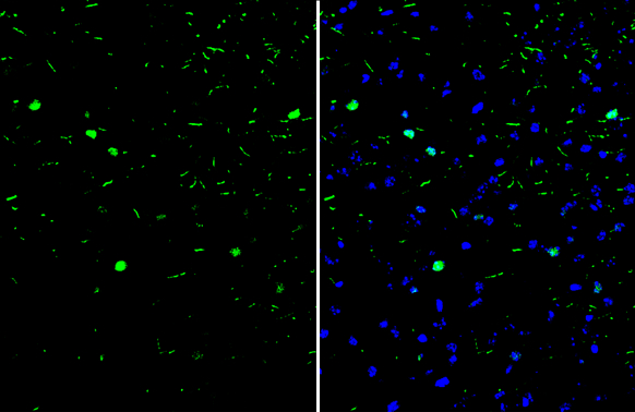
c-Fos antibody detects c-Fos protein by immunohistochemical analysis. Sample: Frozen-sectioned mouse cerebral cortex. Green: c-Fos stained by c-Fos antibody (GTX129846) diluted at 1:250. Blue: Fluoroshield with DAPI (GTX30920).
![c-Fos antibody detects c-Fos protein by immunofluorescent analysis. Sample: DIV9 rat E18 primary cortical neuron cells were fixed in 4% paraformaldehyde at RT for 15 min. Green: c-Fos stained by c-Fos antibody (GTX129846) diluted at 1:500. Red: beta Tubulin 3/ Tuj1, stained by beta Tubulin 3/ Tuj1 antibody [GT11710] (GTX631836) diluted at 1:500. Blue: Fluoroshield with DAPI (GTX30920).](https://www.genetex.com/upload/website/prouct_img/normal/GTX129846/GTX129846_42256_20181115_ICC_IF_R_w_23060523_829.webp "c-Fos antibody detects c-Fos protein by immunofluorescent analysis. Sample: DIV9 rat E18 primary cortical neuron cells were fixed in 4% paraformaldehyde at RT for 15 min. Green: c-Fos stained by c-Fos antibody (GTX129846) diluted at 1:500. Red: beta Tubulin 3/ Tuj1, stained by beta Tubulin 3/ Tuj1 antibody [GT11710] (GTX631836) diluted at 1:500. Blue: Fluoroshield with DAPI (GTX30920).")
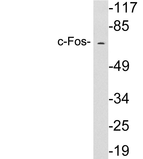
 and treated (+) MCF-7 whole cell extracts (30 μg) were separated by 7.5% SDS-PAGE, and the membrane was blotted with c-Fos antibody (GTX129846) diluted at 1:1000.")
 diluted at 1:500. Red: phalloidin, a cytoskeleton marker, diluted at 1:50. Scale bar = 10 μm.")

c-Fos antibody detects c-Fos protein by immunohistochemical analysis. Sample: Frozen-sectioned mouse cerebral cortex. Green: c-Fos stained by c-Fos antibody (GTX129846) diluted at 1:250. Blue: Fluoroshield with DAPI (GTX30920).
c-Fos antibody
GTX129846
ApplicationsImmunoFluorescence, Western Blot, ImmunoCytoChemistry, ImmunoHistoChemistry, ImmunoHistoChemistry Frozen
Product group Antibodies
ReactivityHuman, Mouse, Rat
TargetFOS
Overview
- SupplierGeneTex
- Product Namec-Fos antibody
- Delivery Days Customer9
- Application Supplier NoteWB: 1:500-1:3000. ICC/IF: 1:100-1:1000. IHC-Fr: 1:100-1:1000. *Optimal dilutions/concentrations should be determined by the researcher.Not tested in other applications.
- ApplicationsImmunoFluorescence, Western Blot, ImmunoCytoChemistry, ImmunoHistoChemistry, ImmunoHistoChemistry Frozen
- CertificationResearch Use Only
- ClonalityPolyclonal
- Concentration0.1 mg/ml
- ConjugateUnconjugated
- Gene ID2353
- Target nameFOS
- Target descriptionFos proto-oncogene, AP-1 transcription factor subunit
- Target synonymsAP-1, C-FOS, p55, protein c-Fos, FBJ murine osteosarcoma viral (v-fos) oncogene homolog (oncogene FOS), FBJ murine osteosarcoma viral oncogene homolog, Fos proto-oncogene, AP-1 trancription factor subunit, G0/G1 switch regulatory protein 7, activator protein 1, cellular oncogene c-fos, proto-oncogene c-Fos, transcription factor AP-1 subunit c-Fos
- HostRabbit
- IsotypeIgG
- Protein IDP01100
- Protein NameProtein c-Fos
- Scientific DescriptionThe Fos gene family consists of 4 members: FOS, FOSB, FOSL1, and FOSL2. These genes encode leucine zipper proteins that can dimerize with proteins of the JUN family, thereby forming the transcription factor complex AP-1. As such, the FOS proteins have been implicated as regulators of cell proliferation, differentiation, and transformation. In some cases, expression of the FOS gene has also been associated with apoptotic cell death. [provided by RefSeq]
- ReactivityHuman, Mouse, Rat
- Storage Instruction-20°C or -80°C,2°C to 8°C
- UNSPSC12352203
References
- Zhu X, Yan F, Liu L, et al. ZEB1 regulates bone metabolism in osteoporotic rats through inducing POLDIP2 transcription. J Orthop Surg Res. 2022,17(1):423. doi: 10.1186/s13018-022-03312-0Read this paper
- Chaney R, Garnier P, Quirié A, et al. Region-Dependent Increase of Cerebral Blood Flow During Electrically Induced Contraction of the Hindlimbs in Rats. Front Physiol. 2022,13:811118. doi: 10.3389/fphys.2022.811118Read this paper
- Mutti V, Bono F, Tomasoni Z, et al. Structural Plasticity of Dopaminergic Neurons Requires the Activation of the D3R-nAChR Heteromer and the PI3K-ERK1/2/Akt-Induced Expression of c-Fos and p70S6K Signaling Pathway. Mol Neurobiol. 2022,59(4):2129-2149. doi: 10.1007/s12035-022-02748-zRead this paper
- Panayotis N, Freund PA, Marvaldi L, et al. β-sitosterol reduces anxiety and synergizes with established anxiolytic drugs in mice. Cell Rep Med. 2021,2(5):100281. doi: 10.1016/j.xcrm.2021.100281Read this paper
- Chang CH, Liu YC, Sun CY, et al. Regulation of stress-provoked aggressive behavior using endocannabinoids. Neurobiol Stress. 2021,15:100337. doi: 10.1016/j.ynstr.2021.100337Read this paper
- Cheng YH, Chiang EI, Syu JN, et al. Treatment of 13-cis retinoic acid and 1,25-dihydroxyvitamin D3 inhibits TNF-alpha-mediated expression of MMP-9 protein and cell invasion through the suppression of JNK pathway and microRNA 221 in human pancreatic adenocarcinoma cancer cells. PLoS One. 2021,16(3):e0247550. doi: 10.1371/journal.pone.0247550Read this paper
- Zhu S, Yang N, Guan Y, et al. GDF15 promotes glioma stem cell-like phenotype via regulation of ERK1/2-c-Fos-LIF signaling. Cell Death Discov. 2021,7(1):3. doi: 10.1038/s41420-020-00395-8Read this paper
- Pan CH, Chen SY, Wang JY, et al. Sclareol ameliorated ERCC1-mediated cisplatin resistance in A549 human lung adenocarcinoma cells and a murine xenograft tumor model by suppressing AKT-GSK3β-AP1/Snail and JNK-AP1 pathways. Chem Biol Interact. 2020,332:109304. doi: 10.1016/j.cbi.2020.109304Read this paper
- Marco-Manclus P, Paredes RG, Portillo W. Sexual experience with a known male modulates c-Fos expression in response to mating and male pheromone exposure in female mice. Physiol Behav. 2020,222:112906. doi: 10.1016/j.physbeh.2020.112906Read this paper
- Leewananthawet A, Arakawa S, Okano T, et al. Ozone ultrafine bubble water induces the cellular signaling involved in oxidative stress responses in human periodontal ligament fibroblasts. Sci Technol Adv Mater. 2019,20(1):589-598. doi: 10.1080/14686996.2019.1614980Read this paper
Datasheet
Related products

Product group Antibodies
Anti-c-Fos/FOS Antibody Picoband(r)A00297-1-CARRIER-FREE
ApplicationsFlow Cytometry, Western Blot, ELISA
ReactivityHuman
TargetFOS
- SizePrice
Product group Antibodies

Anti-cFos [C2-82]Ab02287-10.0
ApplicationsImmunoFluorescence, Western Blot, ELISA, ImmunoHistoChemistry
ReactivityHuman
TargetFOS
- SizePrice
Product group Antibodies
Anti-FOS Antibody144-00236
ApplicationsWestern Blot, ImmunoHistoChemistry
ReactivityHuman, Mouse
TargetFOS
- SizePrice

Product group Antibodies
Anti-FOS AntibodyAMAB91417
ApplicationsWestern Blot
ReactivityHuman
TargetFOS
- SizePrice
Product group Antibodies
References
c-fos Polyclonal Antibodybs-0469R
ApplicationsImmunoFluorescence, Western Blot, ELISA, ImmunoCytoChemistry, ImmunoHistoChemistry, ImmunoHistoChemistry Frozen, ImmunoHistoChemistry Paraffin
ReactivityBovine, Canine, Equine, Human, Mouse, Porcine, Rabbit, Rat
TargetFOS
- SizePrice
Product group Antibodies
FOS Monoclonal AntibodyCSB-MA080264
ApplicationsWestern Blot, ELISA
ReactivityHuman, Mouse, Rat
TargetFOS
- SizePrice
Product group Antibodies

FOS / c-FOS Antibody (clone AWUE3)LS-C764956
ApplicationsWestern Blot, ImmunoHistoChemistry, ImmunoHistoChemistry Paraffin
ReactivityHuman, Mouse, Rat
TargetFOS
- SizePrice
Product group Antibodies
anti-c-Fos (human), Rabbit Monoclonal (RM374)REV-31-1260-00
ApplicationsWestern Blot, ImmunoHistoChemistry
ReactivityHuman
TargetFOS
- SizePrice
Product group Antibodies
Anti-c-Fos AntibodyA97643
ApplicationsWestern Blot, ELISA
ReactivityHuman, Mouse, Rat
- SizePrice