

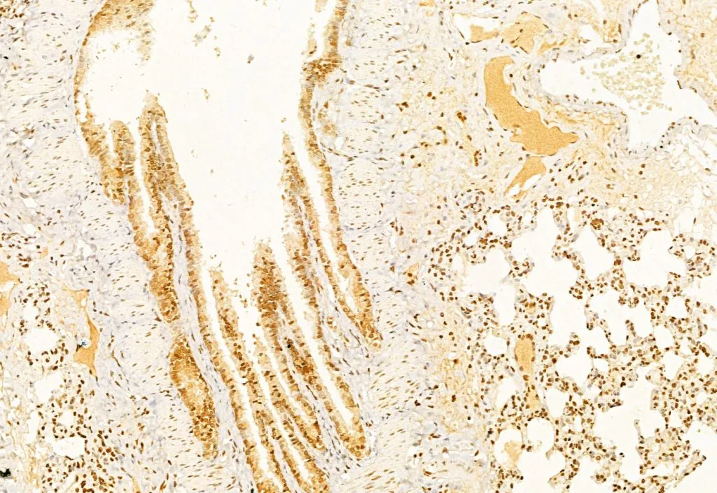
IHC-P analysis of human ehrlich carcimoma tissue using GTX03281 Caspase 3 (cleaved Asp175) antibody. Antigen retrieval : Heat mediated antigen retrieval step in citrate buffer was performed. Dilution : 1:100
 antibody. Lane 1 : HeLa treated with blocking peptide Lane 2 : HeLa (etoposide treated, 25μM 5h) Lane 3 : MCF7 (etoposide treated, 25μM 5h) Lane 4 : Mouse heart tissue lysate Lane 5 : Mouse spleen tissue lysate")
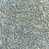
 antibody. Red : Primary antibody Green : beta tubulin Blue : DAPI Permeabilization : 0.1% Triton X-100 Dilution : 1:200")
 antibody. Antigen retrieval : Heat mediated antigen retrieval step in citrate buffer was performed. Dilution : 1:100")
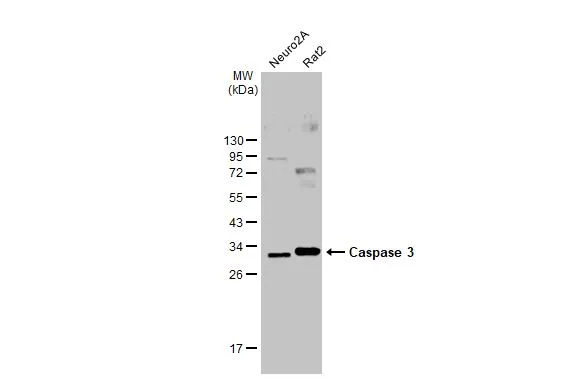
 antibody. The lane on the left was treated with blocking peptide.")
IHC-P analysis of human ehrlich carcimoma tissue using GTX03281 Caspase 3 (cleaved Asp175) antibody. Antigen retrieval : Heat mediated antigen retrieval step in citrate buffer was performed. Dilution : 1:100
Caspase 3 (cleaved Asp175) antibody
GTX03281
ApplicationsImmunoFluorescence, Western Blot, ImmunoCytoChemistry, ImmunoHistoChemistry, ImmunoHistoChemistry Paraffin
Product group Antibodies
ReactivityHuman, Mouse
TargetCASP3
Overview
- SupplierGeneTex
- Product NameCaspase 3 (cleaved Asp175) antibody
- Delivery Days Customer9
- Application Supplier NoteWB: 1:500-1:2000. ICC/IF: 1:100-1:500. IHC-P: 1:50-1:200. *Optimal dilutions/concentrations should be determined by the researcher.Not tested in other applications.
- ApplicationsImmunoFluorescence, Western Blot, ImmunoCytoChemistry, ImmunoHistoChemistry, ImmunoHistoChemistry Paraffin
- CertificationResearch Use Only
- ClonalityPolyclonal
- Concentration1 mg/ml
- ConjugateUnconjugated
- Gene ID836
- Target nameCASP3
- Target descriptioncaspase 3
- Target synonymsCPP32, CPP32B, SCA-1, caspase-3, CASP-3, CPP-32, PARP cleavage protease, SREBP cleavage activity 1, apopain, caspase 3, apoptosis-related cysteine peptidase, caspase 3, apoptosis-related cysteine protease, cysteine protease CPP32, procaspase3, protein Yama
- HostRabbit
- IsotypeIgG
- Protein IDP42574
- Protein NameCaspase-3
- Scientific DescriptionThe protein encoded by this gene is a cysteine-aspartic acid protease that plays a central role in the execution-phase of cell apoptosis. The encoded protein cleaves and inactivates poly(ADP-ribose) polymerase while it cleaves and activates sterol regulatory element binding proteins as well as caspases 6, 7, and 9. This protein itself is processed by caspases 8, 9, and 10. It is the predominant caspase involved in the cleavage of amyloid-beta 4A precursor protein, which is associated with neuronal death in Alzheimers disease. [provided by RefSeq, Aug 2017]
- ReactivityHuman, Mouse
- Storage Instruction-20°C or -80°C,2°C to 8°C
- UNSPSC12352203
References
- Li Y, Wang Z. Interleukin 32 participates in cardiomyocyte-induced oxidative stress, inflammation and apoptosis during hypoxia/reoxygenation via the NOD2/NOX2/MAPK signaling pathway. Exp Ther Med. 2022,24(3):567. doi: 10.3892/etm.2022.11504Read this paper
Datasheet
Related products
Product group Antibodies

Anti-Caspase-3 [4F-6]AB04219-1.1
ApplicationsWestern Blot, ELISA, Neutralisation/Blocking
ReactivityHuman
TargetCASP3
- SizePrice
Product group Antibodies
Anti-CASP3 (138-157aa) Antibody130-10514
ApplicationsELISA
ReactivityHuman
TargetCASP3
- SizePrice

Product group Antibodies
Anti-Caspase-3(p17)/CASP3 Antibody Picoband(r)A00334-2-CARRIER-FREE
ApplicationsFlow Cytometry, ImmunoFluorescence, Western Blot, ImmunoCytoChemistry
ReactivityHuman
TargetCASP3
- SizePrice
Product group Antibodies
References
Caspase 3 antibodyGTX123678
ApplicationsWestern Blot, ImmunoHistoChemistry, ImmunoHistoChemistry Paraffin
ReactivityHuman, Insect, Mouse, Rat
TargetCASP3
- SizePrice
Product group Antibodies
Caspase 3 antibodyGTX04908
ApplicationsImmunoFluorescence, Western Blot, ImmunoCytoChemistry, ImmunoHistoChemistry, ImmunoHistoChemistry Paraffin
ReactivityBovine, Human, Mouse, Rat
TargetCASP3
- SizePrice
Product group Antibodies
References
Caspase 3 antibodyGTX110543
ApplicationsDot Blot, ImmunoPrecipitation, Western Blot, ImmunoHistoChemistry, ImmunoHistoChemistry Frozen, ImmunoHistoChemistry Paraffin
ReactivityHuman, Mouse, Rat
TargetCASP3
- SizePrice
![WB analysis of HeLa cells treated with camptothecin (2μM) lysate using GTX59551 Caspase 3 antibody [C33].](https://www.genetex.com/upload/website/prouct_img/normal/GTX59551/Caspase-3-antibody-C33-GTX59551-WB-1_18121409_343_w_23061123_868.webp)
Product group Antibodies
References
Caspase 3 antibody [C33]GTX59551
ApplicationsWestern Blot
ReactivityHuman, Mouse, Rat
TargetCASP3
- SizePrice
![Wild-type (WT) and Caspase 3 knockout (KO) HeLa cell extracts (30 μg) were separated by 15% SDS-PAGE, and the membrane was blotted with Caspase 3 antibody [HL1370] (GTX636810) diluted at 1:1000. The HRP-conjugated anti-rabbit IgG antibody (GTX213110-01) was used to detect the primary antibody.](https://www.genetex.com/upload/website/prouct_img/normal/GTX636810/GTX636810_44662_20220513_WB_KO_watermark_w_23061202_600.webp)
Product group Antibodies
Caspase 3 antibody [HL1370]GTX636810
ApplicationsWestern Blot, ImmunoHistoChemistry, ImmunoHistoChemistry Paraffin
ReactivityHuman, Rat
TargetCASP3
- SizePrice
Product group Antibodies
References
Caspase 3 antibodyGTX73090
ApplicationsImmunoPrecipitation, Western Blot, ImmunoHistoChemistry, ImmunoHistoChemistry Paraffin
ReactivityHuman, Mouse, Rabbit, Rat
TargetCASP3
- SizePrice