

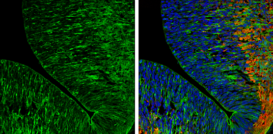
Caspr2 antibody [N2N3] detects Caspr2 protein expression by immunohistochemical analysis. Sample: Frozen sectioned E13.5 Rat brain. Green: Caspr2 protein stained by Caspr2 antibody [N2N3] (GTX109389) diluted at 1:250. Red: beta Tubulin 3/ TUJ1, a mature neuron marker, stained by beta Tubulin 3/ TUJ1 antibody [GT11710] (GTX631836) diluted at 1:500. Blue: Fluoroshield with DAPI (GTX30920).
![Caspr2 antibody [N2N3] detects Caspr2n proteins in embryonic mouse brain by immunohistochemical analysis. Sample: Frozen section of embryonic mouse brain (mE18.5). Red: Caspr2 antibody [N2N3] (GTX109389) diluted at 1:250. Blue: DAPI.](https://www.genetex.com/upload/website/prouct_img/normal/GTX109389/GTX109389_40023_20150703_IHC-Fr_M_w_23060500_960.webp "Caspr2 antibody [N2N3] detects Caspr2n proteins in embryonic mouse brain by immunohistochemical analysis. Sample: Frozen section of embryonic mouse brain (mE18.5). Red: Caspr2 antibody [N2N3] (GTX109389) diluted at 1:250. Blue: DAPI.")
![Caspr2 antibody [N2N3] detects Caspr2 protein by immunofluorescent analysis. Sample: DIV14 rat E18 primary cortical neurons were fixed in 4% paraformaldehyde at RT for 15 min. Green: Caspr2 protein stained by Caspr2 antibody [N2N3] (GTX109389) diluted at 1:500. Red: beta Tubulin 3/ Tuj1, stained by beta Tubulin 3/ Tuj1 antibody [GT1338] (GTX631831) diluted at 1:500. Blue: Fluoroshield with DAPI (GTX30920).](https://www.genetex.com/upload/website/prouct_img/normal/GTX109389/GTX109389_40023_20170719_IFA_R_w_23060500_812.webp "Caspr2 antibody [N2N3] detects Caspr2 protein by immunofluorescent analysis. Sample: DIV14 rat E18 primary cortical neurons were fixed in 4% paraformaldehyde at RT for 15 min. Green: Caspr2 protein stained by Caspr2 antibody [N2N3] (GTX109389) diluted at 1:500. Red: beta Tubulin 3/ Tuj1, stained by beta Tubulin 3/ Tuj1 antibody [GT1338] (GTX631831) diluted at 1:500. Blue: Fluoroshield with DAPI (GTX30920).")
 was separated by 5 % SDS-PAGE, and blotted with Caspr2 antibody (GTX109389) diluted by 1:5000")
![Rat tissue extract (50 μg) was separated by 5% SDS-PAGE, and the membrane was blotted with Caspr2 antibody [N2N3] (GTX109389) diluted at 1:10000. The HRP-conjugated anti-rabbit IgG antibody (GTX213110-01) was used to detect the primary antibody.](https://www.genetex.com/upload/website/prouct_img/normal/GTX109389/GTX109389_41766_20250404_WB_R_brain_25041720_963.webp "Rat tissue extract (50 μg) was separated by 5% SDS-PAGE, and the membrane was blotted with Caspr2 antibody [N2N3] (GTX109389) diluted at 1:10000. The HRP-conjugated anti-rabbit IgG antibody (GTX213110-01) was used to detect the primary antibody.")
![Various tissue extracts (50 μg) were separated by 5% SDS-PAGE, and the membrane was blotted with Caspr2 antibody [N2N3] (GTX109389) diluted at 1:10000. The HRP-conjugated anti-rabbit IgG antibody (GTX213110-01) was used to detect the primary antibody.](https://www.genetex.com/upload/website/prouct_img/normal/GTX109389/GTX109389_41766_20250418_WB_M_tissue_25042420_547.webp "Various tissue extracts (50 μg) were separated by 5% SDS-PAGE, and the membrane was blotted with Caspr2 antibody [N2N3] (GTX109389) diluted at 1:10000. The HRP-conjugated anti-rabbit IgG antibody (GTX213110-01) was used to detect the primary antibody.")
Caspr2 antibody [N2N3] detects Caspr2 protein expression by immunohistochemical analysis. Sample: Frozen sectioned E13.5 Rat brain. Green: Caspr2 protein stained by Caspr2 antibody [N2N3] (GTX109389) diluted at 1:250. Red: beta Tubulin 3/ TUJ1, a mature neuron marker, stained by beta Tubulin 3/ TUJ1 antibody [GT11710] (GTX631836) diluted at 1:500. Blue: Fluoroshield with DAPI (GTX30920).
Caspr2 antibody [N2N3]
GTX109389
ApplicationsImmunoFluorescence, Western Blot, ImmunoCytoChemistry, ImmunoHistoChemistry, ImmunoHistoChemistry Frozen
Product group Antibodies
ReactivityHuman, Mouse, Rat
TargetCNTNAP2
Overview
- SupplierGeneTex
- Product NameCaspr2 antibody [N2N3]
- Delivery Days Customer9
- Application Supplier NoteWB: 1:5000-1:20000. ICC/IF: 1:100-1:1000. IHC-Fr: 1:100-1:1000. *Optimal dilutions/concentrations should be determined by the researcher.Not tested in other applications.
- ApplicationsImmunoFluorescence, Western Blot, ImmunoCytoChemistry, ImmunoHistoChemistry, ImmunoHistoChemistry Frozen
- CertificationResearch Use Only
- ClonalityPolyclonal
- Concentration1 mg/ml
- ConjugateUnconjugated
- Gene ID26047
- Target nameCNTNAP2
- Target descriptioncontactin associated protein 2
- Target synonymsAUTS15, CASPR2, CDFE, NRXN4, PTHSL1, contactin-associated protein-like 2, cell recognition molecule Caspr2, contactin associated protein like 2, homolog of Drosophila neurexin IV
- HostRabbit
- IsotypeIgG
- Protein IDQ9UHC6
- Protein NameContactin-associated protein-like 2
- Scientific DescriptionThis gene encodes a member of the neurexin family which functions in the vertebrate nervous system as cell adhesion molecules and receptors. This protein, like other neurexin proteins, contains epidermal growth factor repeats and laminin G domains. In addition, it includes an F5/8 type C domain, discoidin/neuropilin- and fibrinogen-like domains, thrombospondin N-terminal-like domains and a putative PDZ binding site. This protein is localized at the juxtaparanodes of myelinated axons and associated with potassium channels. It may play a role in the local differentiation of the axon into distinct functional subdomains. This gene encompasses almost 1.5% of chromosome 7 and is one of the largest genes in the human genome. It may represent a positional candidate gene for the DFNB13 form of nonsyndromic deafness. [provided by RefSeq]
- ReactivityHuman, Mouse, Rat
- Storage Instruction-20°C or -80°C,2°C to 8°C
- UNSPSC41116161
Datasheet
Related products

Product group Antibodies
Anti-Caspr2/CNTNAP2 Antibody Picoband(r)A02819-2-CARRIER-FREE
ApplicationsWestern Blot, ELISA
ReactivityHuman, Mouse, Rat
TargetCNTNAP2
- SizePrice

Product group Antibodies
Anti-CNTNAP2 AntibodyHPA002739
ApplicationsWestern Blot, ImmunoHistoChemistry
ReactivityHuman
TargetCNTNAP2
- SizePrice
Product group Antibodies
CNTNAP2 AntibodyCSB-PA887030ESR2HU
ApplicationsELISA
ReactivityHuman
TargetCNTNAP2
- SizePrice
Product group Antibodies

CNTNAP2 / CASPR2 AntibodyLS-C409156
ApplicationsWestern Blot, ImmunoHistoChemistry
ReactivityHuman, Mouse
TargetCNTNAP2
- SizePrice
Product group Antibodies
Anti-CNTNAP2 (Center) Antibody102-27581
ApplicationsWestern Blot, ImmunoHistoChemistry, ImmunoHistoChemistry Paraffin
TargetCNTNAP2
- SizePrice
Product group Antibodies
Caspr2 Polyclonal AntibodyBS-11129R
ApplicationsFlow Cytometry, Western Blot, ELISA
TargetCNTNAP2
- SizePrice