

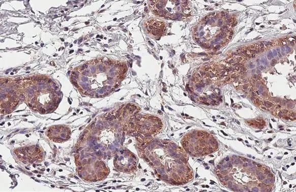
CCR1 antibody [HL2840] detects CCR1 protein at cell membrane and cytoplasm by immunohistochemical analysis. Sample: Paraffin-embedded human breast carcinoma. CCR1 stained by CCR1 antibody [HL2840] (GTX640111) diluted at 1:100. Antigen Retrieval: Citrate buffer, pH 6.0, 15 min
![Non-transfected (–) and transfected (+) boiled and unboiled 293T whole cell extracts (30 μg) were separated by 10% SDS-PAGE, and the membrane was blotted with CCR1 antibody [HL2840] (GTX640111) diluted at 1:5000. The HRP-conjugated anti-rabbit IgG antibody (GTX213110-01) was used to detect the primary antibody.](https://www.genetex.com/upload/website/prouct_img/normal/GTX640111/GTX640111_T-45355_20240607_WB_B_24061301_221.webp "Non-transfected (–) and transfected (+) boiled and unboiled 293T whole cell extracts (30 μg) were separated by 10% SDS-PAGE, and the membrane was blotted with CCR1 antibody [HL2840] (GTX640111) diluted at 1:5000. The HRP-conjugated anti-rabbit IgG antibody (GTX213110-01) was used to detect the primary antibody.")
![Unboiled K562 whole cell and membrane extracts (30 μg) were separated by 10% SDS-PAGE, and the membrane was blotted with CCR1 antibody [HL2840] (GTX640111) diluted at 1:1000. The HRP-conjugated anti-rabbit IgG antibody (GTX213110-01) was used to detect the primary antibody. (WCE: whole cell extract; ME: membrane extract)](https://www.genetex.com/upload/website/prouct_img/normal/GTX640111/GTX640111_45432_20240913_WB_Fraction_24091901_727.webp "Unboiled K562 whole cell and membrane extracts (30 μg) were separated by 10% SDS-PAGE, and the membrane was blotted with CCR1 antibody [HL2840] (GTX640111) diluted at 1:1000. The HRP-conjugated anti-rabbit IgG antibody (GTX213110-01) was used to detect the primary antibody. (WCE: whole cell extract; ME: membrane extract)")
![CCR1 antibody [HL2840] detects CCR1 protein by immunohistochemical analysis. Sample: Paraffin-embedded human tissues. CCR1 stained by CCR1 antibody [HL2840] (GTX640111) diluted at 1:100. Antigen Retrieval: Citrate buffer, pH 6.0, 15 min Corresponding RNA expression data for the same tissues are based on NCBI.](https://www.genetex.com/upload/website/prouct_img/normal/GTX640111/GTX640111_45432_20241113_IHC-P_multiple_RPKM_24111918_487.webp "CCR1 antibody [HL2840] detects CCR1 protein by immunohistochemical analysis. Sample: Paraffin-embedded human tissues. CCR1 stained by CCR1 antibody [HL2840] (GTX640111) diluted at 1:100. Antigen Retrieval: Citrate buffer, pH 6.0, 15 min Corresponding RNA expression data for the same tissues are based on NCBI.")
![CCR1 antibody [HL2840] detects CCR1 protein by immunofluorescent analysis. Sample: THP-1 cells were fixed in 4% paraformaldehyde at RT for 15 min. Green: CCR1 stained by CCR1 antibody [HL2840] (GTX640111) diluted at 1:100. Blue: Fluoroshield with DAPI (GTX30920).](https://www.genetex.com/upload/website/prouct_img/normal/GTX640111/GTX640111_45432_20241122_ICC_IF_24120522_100.webp "CCR1 antibody [HL2840] detects CCR1 protein by immunofluorescent analysis. Sample: THP-1 cells were fixed in 4% paraformaldehyde at RT for 15 min. Green: CCR1 stained by CCR1 antibody [HL2840] (GTX640111) diluted at 1:100. Blue: Fluoroshield with DAPI (GTX30920).")
![Whole cell extract (30 μg) was separated by 10% SDS-PAGE, and the membrane was blotted with CCR1 antibody [HL2840] (GTX640111) diluted at 1:1000. The HRP-conjugated anti-rabbit IgG antibody (GTX213110-01) was used to detect the primary antibody, and the signal was developed with Trident ECL plus-Enhanced.](https://www.genetex.com/upload/website/prouct_img/normal/GTX640111/GTX640111_45432_20250131_WB_C_25020422_829.webp "Whole cell extract (30 μg) was separated by 10% SDS-PAGE, and the membrane was blotted with CCR1 antibody [HL2840] (GTX640111) diluted at 1:1000. The HRP-conjugated anti-rabbit IgG antibody (GTX213110-01) was used to detect the primary antibody, and the signal was developed with Trident ECL plus-Enhanced.")
![Non-transfected (–) and transfected (+) unboiled K562 whole cell extracts (50 μg) were separated by 10% SDS-PAGE, and the membrane was blotted with CCR1 antibody [HL2840] (GTX640111) diluted at 1:500. The HRP-conjugated anti-rabbit IgG antibody (GTX213110-01) was used to detect the primary antibody, and the signal was developed with Trident femto Western HRP Substrate.](https://www.genetex.com/upload/website/prouct_img/normal/GTX640111/GTX640111_45432_20251107_WB_shRNA_watermark_25111401_235.webp "Non-transfected (–) and transfected (+) unboiled K562 whole cell extracts (50 μg) were separated by 10% SDS-PAGE, and the membrane was blotted with CCR1 antibody [HL2840] (GTX640111) diluted at 1:500. The HRP-conjugated anti-rabbit IgG antibody (GTX213110-01) was used to detect the primary antibody, and the signal was developed with Trident femto Western HRP Substrate.")
CCR1 antibody [HL2840] detects CCR1 protein at cell membrane and cytoplasm by immunohistochemical analysis. Sample: Paraffin-embedded human breast carcinoma. CCR1 stained by CCR1 antibody [HL2840] (GTX640111) diluted at 1:100. Antigen Retrieval: Citrate buffer, pH 6.0, 15 min
CCR1 antibody [HL2840]
GTX640111
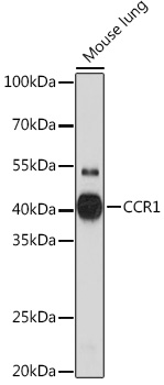
ApplicationsImmunoFluorescence, Western Blot, ImmunoCytoChemistry, ImmunoHistoChemistry, ImmunoHistoChemistry Paraffin
Product group Antibodies
ReactivityFeline, Human
TargetCCR1
Overview
- SupplierGeneTex
- Product NameCCR1 antibody [HL2840]
- Delivery Days Customer7
- Application Supplier NoteIHC-P: 1:100-1:1000. *Optimal dilutions/concentrations should be determined by the researcher.Not tested in other applications.
- ApplicationsImmunoFluorescence, Western Blot, ImmunoCytoChemistry, ImmunoHistoChemistry, ImmunoHistoChemistry Paraffin
- CertificationResearch Use Only
- ClonalityMonoclonal
- Clone IDHL2840
- Concentration1 mg/ml
- ConjugateUnconjugated
- Gene ID1230
- Target nameCCR1
- Target descriptionC-C motif chemokine receptor 1
- Target synonymsCD191, CKR-1, CKR1, CMKBR1, HM145, MIP1aR, SCYAR1, C-C chemokine receptor type 1, C-C CKR-1, CC-CKR-1, CCR-1, LD78 receptor, MIP-1alpha-R, RANTES receptor, RANTES-R, chemokine (C-C motif) receptor 1, macrophage inflammatory protein 1-alpha receptor
- HostRabbit
- IsotypeIgG
- Protein IDP32246
- Protein NameC-C chemokine receptor type 1
- Scientific DescriptionThis gene encodes a member of the beta chemokine receptor family, which is predicted to be a seven transmembrane protein similar to G protein-coupled receptors. The ligands of this receptor include macrophage inflammatory protein 1 alpha (MIP-1 alpha), regulated on activation normal T expressed and secreted protein (RANTES), monocyte chemoattractant protein 3 (MCP-3), and myeloid progenitor inhibitory factor-1 (MPIF-1). Chemokines and their receptors mediated signal transduction are critical for the recruitment of effector immune cells to the site of inflammation. Knockout studies of the mouse homolog suggested the roles of this gene in host protection from inflammatory response, and susceptibility to virus and parasite. This gene and other chemokine receptor genes, including CCR2, CCRL2, CCR3, CCR5 and CCXCR1, are found to form a gene cluster on chromosome 3p. [provided by RefSeq, Jul 2008]
- ReactivityFeline, Human
- Storage Instruction-20°C or -80°C,2°C to 8°C
- UNSPSC41116161
Datasheet
Related products
Product group Antibodies
Anti-CCR1 AntibodyA307305
ApplicationsImmunoFluorescence, Western Blot, ImmunoCytoChemistry, ImmunoHistoChemistry
ReactivityMouse, Rat
- SizePrice
Product group Antibodies
ApplicationsFlow Cytometry
ReactivityHuman
TargetCCR1
- SizePrice

Product group Antibodies
Anti-CKR-1 CCR1 AntibodyA01896
ApplicationsImmunoFluorescence, ELISA, ImmunoHistoChemistry
ReactivityHuman, Mouse
TargetCCR1
- SizePrice
Product group Antibodies
Ccr1 Polyclonal AntibodyCAC08295
ApplicationsImmunoFluorescence, ELISA, ImmunoHistoChemistry
TargetCCR1
- SizePrice
Product group Antibodies
CCR1 AntibodyCSB-PA004839LA01HU
ApplicationsImmunoFluorescence, ELISA, ImmunoHistoChemistry
ReactivityHuman
TargetCCR1
- SizePrice
Product group Antibodies
CCR1 antibodyGTX13240
ApplicationsFlow Cytometry, Western Blot, ELISA, ImmunoHistoChemistry
ReactivityHuman
TargetCCR1
- SizePrice
Product group Antibodies
CCR1 antibodyGTX70509
ApplicationsImmunoHistoChemistry, ImmunoHistoChemistry Paraffin
ReactivityHuman
TargetCCR1
- SizePrice
Product group Antibodies
CCR1 antibody [7B17]GTX52512
ApplicationsFlow Cytometry, ImmunoHistoChemistry, ImmunoHistoChemistry Paraffin
ReactivityHuman
TargetCCR1
- SizePrice
![CCR1 antibody [HL3023] detects CCR1 protein by immunofluorescent analysis. Sample: Mock and transfected 293T cells were fixed in ice-cold MeOH for 5 min. Green: CCR1 stained by CCR1 antibody [HL3023] (GTX640436) diluted at 1:500. Blue: Fluoroshield with DAPI (GTX30920).](https://www.genetex.com/upload/website/prouct_img/normal/GTX640436/GTX640436_T-45425_20240628_ICC_IF_B_24080622_951.webp)
Product group Antibodies
CCR1 antibody [HL3023]GTX640436
ApplicationsImmunoFluorescence, ImmunoCytoChemistry, ImmunoHistoChemistry, ImmunoHistoChemistry Paraffin
ReactivityHuman, Mouse
TargetCCR1
- SizePrice
![Non-transfected (–) and transfected (+) boiled and unboiled 293T whole cell extracts (30 μg) were separated by 10% SDS-PAGE, and the membrane was blotted with CCR1 antibody [HL3034] (GTX640472) diluted at 1:5000. The HRP-conjugated anti-rabbit IgG antibody (GTX213110-01) was used to detect the primary antibody.](https://www.genetex.com/upload/website/prouct_img/normal/GTX640472/GTX640472_T-45432_20240607_WB_B_24061301_834.webp)
Product group Antibodies
CCR1 antibody [HL3034]GTX640472
ApplicationsImmunoFluorescence, Western Blot, ImmunoCytoChemistry, ImmunoHistoChemistry, ImmunoHistoChemistry Paraffin
ReactivityFeline, Human
TargetCCR1
- SizePrice