

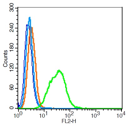
HepG2 cells probed with CD63/MLA1 Polyclonal Antibody, Unconjugated (bs-1523R) at 1:100 for 30 minutes followed by incubation with a conjugated secondary (PE Conjugated) (green) for 30 minutes compared to control cells (blue), secondary only (light blue) and isotype control (orange).
 Polyclonal Antibody, Unconjugated (Catalog #) at 1:300 overnight at 4˚C. Followed by a conjugated secondary antibody at 1:5000 for 90 min at 37˚C.")
 were fixed with 4% PFA for 10min at room temperature,permeabilized with 90% ice-cold methanol for 20 min at -20℃, and incubated in 5% BSA blocking buffer for 30 min at room temperature. Cells were then stained with CD63/MLA1 Polyclonal Antibody(bs-1523R)at 1:50 dilution in blocking buffer and incubated for 30 min at room temperature, washed twice with 2% BSA in PBS, followed by secondary antibody(blue) incubation for 40 min at room temperature. Acquisitions of 20,000 events were performed. Cells stained with primary antibody (green), and isotype control (orange).")
HepG2 cells probed with CD63/MLA1 Polyclonal Antibody, Unconjugated (bs-1523R) at 1:100 for 30 minutes followed by incubation with a conjugated secondary (PE Conjugated) (green) for 30 minutes compared to control cells (blue), secondary only (light blue) and isotype control (orange).
CD63 Polyclonal Antibody
BS-1523R
ApplicationsFlow Cytometry, ImmunoFluorescence, Western Blot, ELISA, ImmunoCytoChemistry, ImmunoHistoChemistry, ImmunoHistoChemistry Frozen, ImmunoHistoChemistry Paraffin
Product group Antibodies
ReactivityHuman
TargetCD63
Overview
- SupplierBioss
- Product NameCD63 Polyclonal Antibody
- Delivery Days Customer16
- ApplicationsFlow Cytometry, ImmunoFluorescence, Western Blot, ELISA, ImmunoCytoChemistry, ImmunoHistoChemistry, ImmunoHistoChemistry Frozen, ImmunoHistoChemistry Paraffin
- Applications SupplierWB(1:300-5000), ELISA(1:500-1000), FCM(1:20-100), IHC-P(1:200-400), IHC-F(1:100-500), IF(IHC-P)(1:50-200), IF(IHC-F)(1:50-200), IF(ICC)(1:50-200)
- CertificationResearch Use Only
- ClonalityPolyclonal
- Concentration1 ug/ul
- ConjugateUnconjugated
- Gene ID967
- Target nameCD63
- Target descriptionCD63 molecule
- Target synonymsAD1, HOP-26, ME491, MLA1, OMA81H, Pltgp40, TSPAN30, CD63 antigen, AD1 antigen, CD63 antigen (melanoma 1 antigen), granulophysin, limp1, melanoma-associated antigen ME491, melanoma-associated antigen MLA1, ocular melanoma-associated antigen, tetraspanin-30, tspan-30
- HostRabbit
- IsotypeIgG
- Protein IDP08962
- Protein NameCD63 antigen
- ReactivityHuman
- Storage Instruction-20°C
- UNSPSC41116161
References
- Glioblastoma Stem Cell-Derived Exosomes Enhance Stemness and Tumorigenicity of Glioma Cells by Transferring Notch1 Protein. Sun Z et al., 2020 Jul, Cell Mol NeurobiolRead this paper
- Single-Molecule Co-Immunoprecipitation Reveals Functional Inheritance of EGFRs in Extracellular Vesicles. Sung MS et al., 2018 Oct, SmallRead this paper
- Osteoclast-derived microRNA-containing exosomes selectively inhibit osteoblast activity. Sun W et al., 2016, Cell DiscovRead this paper
- Highly potent intracellular membrane-associated Abeta seeds. Marzesco AM et al., 2016 Jun 17, Sci RepRead this paper
Datasheet
Related products
Product group Antibodies

Anti-CD63 [MOF11]Ab00388-1.1
ApplicationsFlow Cytometry
ReactivityHuman
TargetCD63
- SizePrice

Product group Antibodies
Anti-CD63 AntibodyA101276
ApplicationsELISA, ImmunoHistoChemistry
ReactivityHuman
- SizePrice
Product group Antibodies
CD63 AntibodyCSB-PA006039
ApplicationsWestern Blot, ELISA
ReactivityHuman
TargetCD63
- SizePrice
Product group Antibodies
ApplicationsFlow Cytometry
TargetCD63
- SizePrice
Product group Antibodies

ApplicationsImmunoFluorescence, ELISA, ImmunoHistoChemistry, ImmunoHistoChemistry Frozen, ImmunoHistoChemistry Paraffin
ReactivityHuman
TargetCD63
- SizePrice
Product group Antibodies
CD63 antibodyGTX132953
ApplicationsWestern Blot, ImmunoHistoChemistry, ImmunoHistoChemistry Paraffin
ReactivityHuman
TargetCD63
- SizePrice
Product group Antibodies

CD63 Antibody (Preservative Free)LS-C342960
ApplicationsWestern Blot, ELISA
ReactivityHuman
TargetCD63
- SizePrice

Product group Antibodies
Anti-CD63 AntibodyHPA010088
ApplicationsImmunoHistoChemistry
ReactivityHuman
TargetCD63
- SizePrice