

CD8(C8/468), Biotin conjugate, 0.1mg/mL [26628-22-8]
BNCB0468
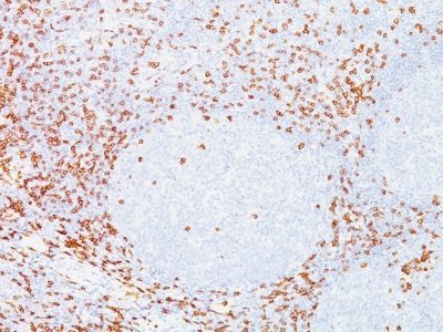
ApplicationsImmunoFluorescence, ImmunoHistoChemistry, ImmunoHistoChemistry Paraffin
Product group Antibodies
ReactivityBovine, Human, Mouse
TargetCD8A
Overview
- SupplierBiotium
- Product NameCD8(C8/468), Biotin conjugate, 0.1mg/mL [26628-22-8]
- Delivery Days Customer9
- ApplicationsImmunoFluorescence, ImmunoHistoChemistry, ImmunoHistoChemistry Paraffin
- CAS Number26628-22-8
- CertificationResearch Use Only
- ClonalityMonoclonal
- Clone IDC8/468
- Concentration0.1 mg/ml
- ConjugateBiotin
- Gene ID925
- Target nameCD8A
- Target descriptionCD8 subunit alpha
- Target synonymsCD8, CD8alpha, IMD116, Leu2, p32, T-cell surface glycoprotein CD8 alpha chain, CD8 antigen, alpha polypeptide (p32), CD8a molecule, Leu2 T-lymphocyte antigen, OKT8 T-cell antigen, T cell co-receptor, T-cell antigen Leu2, T-lymphocyte differentiation antigen T8/Leu-2, T8 T-cell antigen
- HostMouse
- IsotypeIgG1
- Protein IDP01732
- Protein NameT-cell surface glycoprotein CD8 alpha chain
- Scientific DescriptionCD8 is a cell surface receptor expressed either as a heterodimer with the CD8 beta chain (CD8 alpha/beta) or as a homodimer (CD8 alpha/alpha). A majority of thymocytes and a subpopulation of mature T cells and NK cells express CD8a. CD8 binds to MHC class 1 and through its association with protein tyrosine kinase p56lck plays a role in T cell development and activation of mature T cells. For mature T-cells, CD4 and CD8 are mutually exclusive, so anti-CD8, generally used in conjunction with anti-CD4. It is a useful marker for distinguishing helper/inducer T-lymphocytes, and most peripheral T-cell lymphomas are CD4 /CD8-. Anaplastic large cell lymphoma is usually CD4 and CD8-, and in T-lymphoblastic lymphoma/leukemia, CD4 and CD8 are often co-expressed. CD8 is also found in littoral cell angioma of the spleen.Primary antibodies are available purified, or with a selection of fluorescent CF® Dyes and other labels. CF® Dyes offer exceptional brightness and photostability. Note: Conjugates of blue fluorescent dyes like CF®405S and CF®405M are not recommended for detecting low abundance targets, because blue dyes have lower fluorescence and can give higher non-specific background than other dye colors.
- SourceAnimal
- ReactivityBovine, Human, Mouse
- Storage Instruction2°C to 8°C,RT
- UNSPSC41116161
MSDS
Related products
Product group Antibodies
Anti-CD8A AntibodyA28543
ApplicationsWestern Blot
ReactivityHuman, Mouse, Rat
- SizePrice
Product group Antibodies

Anti-CD8 [UCHT4]Ab00694-2.0
ApplicationsFlow Cytometry, ImmunoPrecipitation, ImmunoHistoChemistry, Neutralisation/Blocking
ReactivityHuman, Primate
TargetCD8A
- SizePrice

Product group Antibodies
ApplicationsImmunoCytoChemistry
ReactivityHuman
TargetCD8A
- SizePrice
Product group Antibodies
Anti-CD8A Antibody144-62144
ApplicationsWestern Blot, ImmunoHistoChemistry
ReactivityHuman, Mouse, Rat
TargetCD8A
- SizePrice

Product group Antibodies
Anti-CD8A AntibodyAMAB90883
ApplicationsImmunoHistoChemistry
ReactivityHuman
TargetCD8A
- SizePrice
Product group Antibodies

ApplicationsFlow Cytometry
ReactivityHuman
TargetCD8A
- SizePrice
Product group Antibodies
Cd8A Polyclonal AntibodyCAC07494
ApplicationsELISA, ImmunoHistoChemistry
TargetCD8A
- SizePrice
Product group Antibodies
CD8A AntibodyCSB-PA004966LA01HU
ApplicationsELISA, ImmunoHistoChemistry
ReactivityHuman
TargetCD8A
- SizePrice
Product group Antibodies
References
CD8 Polyclonal AntibodyBS-4790R
ApplicationsFlow Cytometry, ImmunoFluorescence, Western Blot, ELISA, ImmunoCytoChemistry, ImmunoHistoChemistry, ImmunoHistoChemistry Frozen, ImmunoHistoChemistry Paraffin
ReactivityHuman
TargetCD8A
- SizePrice
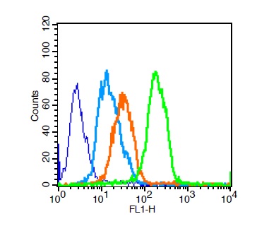
![FACS analysis of human peripheral blood lymphocytes using GTX01467-06 CD8 alpha antibody [RPA-T8] (FITC). Solid lone : primary antibody Dashed line : isotype control antibody amount : 1 μg (5 μl)](https://www.genetex.com/upload/website/prouct_img/normal/GTX01467-06/GTX01467-06_20200428_FACS72_w_23053121_484.webp)
Product group Antibodies
CD8 alpha antibody [RPA-T8] (FITC)GTX01467-06
ApplicationsFlow Cytometry
ReactivityHuman
TargetCD8A
- SizePrice