

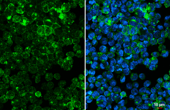
CD97 antibody [HL1924] detects CD97 protein at cell membrane and cytoplasmic vesicles by immunofluorescent analysis. Sample: U937 cells were fixed in ice-cold MeOH for 5 min. Green: CD97 stained by CD97 antibody [HL1924] (GTX637673) diluted at 1:500. Blue: Fluoroshield with DAPI (GTX30920).
![Non-transfected (–) and transfected (+) Boiled and unboiled 293T whole cell extracts (30 μg) were separated by 7.5% SDS-PAGE, and the membrane was blotted with CD97 antibody [HL1924] (GTX637673) diluted at 1:1000. The HRP-conjugated anti-rabbit IgG antibody (GTX213110-01) was used to detect the primary antibody.](https://www.genetex.com/upload/website/prouct_img/normal/GTX637673/GTX637673_45166_20230915_WB_B_23091901_981.webp "Non-transfected (–) and transfected (+) Boiled and unboiled 293T whole cell extracts (30 μg) were separated by 7.5% SDS-PAGE, and the membrane was blotted with CD97 antibody [HL1924] (GTX637673) diluted at 1:1000. The HRP-conjugated anti-rabbit IgG antibody (GTX213110-01) was used to detect the primary antibody.")
![Whole cell extract (30 μg) was separated by 7.5% SDS-PAGE, and the membrane was blotted with CD97 antibody [HL1924] (GTX637673) diluted at 1:1000. The HRP-conjugated anti-rabbit IgG antibody (GTX213110-01) was used to detect the primary antibody, and the signal was developed with Trident ECL plus-Enhanced.](https://www.genetex.com/upload/website/prouct_img/normal/GTX637673/GTX637673_45166_20230922_WB_D_23100118_388.webp "Whole cell extract (30 μg) was separated by 7.5% SDS-PAGE, and the membrane was blotted with CD97 antibody [HL1924] (GTX637673) diluted at 1:1000. The HRP-conjugated anti-rabbit IgG antibody (GTX213110-01) was used to detect the primary antibody, and the signal was developed with Trident ECL plus-Enhanced.")
![Boiled and unboiled wild-type (WT) and ADGRE5 knockout (KO) HeLa cell extracts (30 μg) were separated by 7.5% SDS-PAGE, and the membrane was blotted with CD97 antibody [HL1924] (GTX637673) diluted at 1:1000. The HRP-conjugated anti-rabbit IgG antibody (GTX213110-01) was used to detect the primary antibody.](https://www.genetex.com/upload/website/prouct_img/normal/GTX637673/GTX637673_T-44837_20231006_WB_KO_watermark_23102401_836.webp "Boiled and unboiled wild-type (WT) and ADGRE5 knockout (KO) HeLa cell extracts (30 μg) were separated by 7.5% SDS-PAGE, and the membrane was blotted with CD97 antibody [HL1924] (GTX637673) diluted at 1:1000. The HRP-conjugated anti-rabbit IgG antibody (GTX213110-01) was used to detect the primary antibody.")
![Various whole cell extracts (30 μg) were separated by 7.5% SDS-PAGE, and the membrane was blotted with CD97 antibody [HL1924] (GTX637673) diluted at 1:500. The HRP-conjugated anti-rabbit IgG antibody (GTX213110-01) was used to detect the primary antibody. Corresponding RNA expression data for the same cell lines are based on Human Protein Atlas program.](https://www.genetex.com/upload/website/prouct_img/normal/GTX637673/GTX637673_45166_20240112_WB_TPM_watermark_24011618_239.webp "Various whole cell extracts (30 μg) were separated by 7.5% SDS-PAGE, and the membrane was blotted with CD97 antibody [HL1924] (GTX637673) diluted at 1:500. The HRP-conjugated anti-rabbit IgG antibody (GTX213110-01) was used to detect the primary antibody. Corresponding RNA expression data for the same cell lines are based on Human Protein Atlas program.")
![CD97 antibody [HL1924] detects CD97 protein by immunofluorescent analysis. Sample: Mock and transfected 293T cells were fixed in MeOH. Green: CD97 stained by CD97 antibody [HL1924] (GTX637673) diluted at 1:500. Blue: Fluoroshield with DAPI (GTX30920).](https://www.genetex.com/upload/website/prouct_img/normal/GTX637673/GTX637673_45166_20231229_ICC_IF_B_24011618_433.webp "CD97 antibody [HL1924] detects CD97 protein by immunofluorescent analysis. Sample: Mock and transfected 293T cells were fixed in MeOH. Green: CD97 stained by CD97 antibody [HL1924] (GTX637673) diluted at 1:500. Blue: Fluoroshield with DAPI (GTX30920).")
![Unboiled non-transfected (–) and transfected (+) Expi293 whole cell extracts (30 μg) were separated by 7.5% SDS-PAGE, and the membrane was blotted with CD97 antibody [HL1924] (GTX637673) diluted at 1:5000. The HRP-conjugated anti-rabbit IgG antibody (GTX213110-01) was used to detect the primary antibody.](https://www.genetex.com/upload/website/prouct_img/normal/GTX637673/GTX637673_45166_20250515_WB_multiple_B_25052202_902.webp "Unboiled non-transfected (–) and transfected (+) Expi293 whole cell extracts (30 μg) were separated by 7.5% SDS-PAGE, and the membrane was blotted with CD97 antibody [HL1924] (GTX637673) diluted at 1:5000. The HRP-conjugated anti-rabbit IgG antibody (GTX213110-01) was used to detect the primary antibody.")
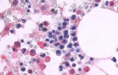
CD97 antibody [HL1924] detects CD97 protein at cell membrane and cytoplasmic vesicles by immunofluorescent analysis. Sample: U937 cells were fixed in ice-cold MeOH for 5 min. Green: CD97 stained by CD97 antibody [HL1924] (GTX637673) diluted at 1:500. Blue: Fluoroshield with DAPI (GTX30920).
CD97 antibody [HL1924]
GTX637673
ApplicationsImmunoFluorescence, Western Blot, ImmunoCytoChemistry
Product group Antibodies
ReactivityCanine, Human
TargetADGRE5
Overview
- SupplierGeneTex
- Product NameCD97 antibody [HL1924]
- Delivery Days Customer9
- Application Supplier NoteWB: 1:500-1:3000. *Optimal dilutions/concentrations should be determined by the researcher.Not tested in other applications.
- ApplicationsImmunoFluorescence, Western Blot, ImmunoCytoChemistry
- CertificationResearch Use Only
- ClonalityMonoclonal
- Clone IDHL1924
- Concentration1 mg/ml
- ConjugateUnconjugated
- Gene ID976
- Target nameADGRE5
- Target descriptionadhesion G protein-coupled receptor E5
- Target synonymsCD97, TM7LN1, adhesion G protein-coupled receptor E5, CD97 molecule, leukocyte antigen CD97, seven transmembrane helix receptor, seven-span transmembrane protein, seven-transmembrane, heterodimeric receptor associated with inflammation
- HostRabbit
- IsotypeIgG
- Protein IDP48960
- Protein NameAdhesion G protein-coupled receptor E5
- Scientific DescriptionThis gene encodes a member of the EGF-TM7 subfamily of adhesion G protein-coupled receptors, which mediate cell-cell interactions. These proteins are cleaved by self-catalytic proteolysis into a large extracellular subunit and seven-span transmembrane subunit, which associate at the cell surface as a receptor complex. The encoded protein may play a role in cell adhesion as well as leukocyte recruitment, activation and migration, and contains multiple extracellular EGF-like repeats which mediate binding to chondroitin sulfate and the cell surface complement regulatory protein CD55. Expression of this gene may play a role in the progression of several types of cancer. Alternatively spliced transcript variants encoding multiple isoforms with 3 to 5 EGF-like repeats have been observed for this gene. This gene is found in a cluster with other EGF-TM7 genes on the short arm of chromosome 19. [provided by RefSeq, Jun 2011]
- ReactivityCanine, Human
- Storage Instruction-20°C or -80°C,2°C to 8°C
- UNSPSC41116161
Datasheet
Related products

Product group Antibodies
Anti-ADGRE5 Antibody Picoband(r)A30392-2-CARRIER-FREE
ApplicationsFlow Cytometry, Western Blot, ELISA
ReactivityHuman
TargetADGRE5
- SizePrice
Product group Antibodies
Anti-CD97 Antibody144-03780
ApplicationsWestern Blot, ImmunoHistoChemistry
ReactivityHuman, Mouse, Rat
TargetADGRE5
- SizePrice
Product group Antibodies
Anti-CD97 AntibodyA14462
ApplicationsWestern Blot
ReactivityHuman, Mouse, Rat
- SizePrice
Product group Antibodies

ADGRE5 / CD97 AntibodyLS-C761072
ApplicationsWestern Blot
ReactivityHuman, Rat
TargetADGRE5
- SizePrice
Product group Antibodies
CD97 Polyclonal AntibodyBS-2522R
ApplicationsFlow Cytometry, ImmunoFluorescence, ELISA, ImmunoCytoChemistry, ImmunoHistoChemistry, ImmunoHistoChemistry Frozen, ImmunoHistoChemistry Paraffin
ReactivityBovine, Canine, Human, Mouse, Porcine, Rat
TargetADGRE5
- SizePrice
Product group Antibodies
ApplicationsImmunoPrecipitation, Western Blot, ImmunoCytoChemistry, ImmunoHistoChemistry
ReactivityPorcine
TargetADGRE5
- SizePrice
Product group Antibodies
CD97 AntibodyCSB-PA004972ESR2HU
ApplicationsWestern Blot, ELISA, ImmunoHistoChemistry
ReactivityHuman
TargetADGRE5
- SizePrice
Product group Antibodies
CD97 antibodyGTX70829
ApplicationsImmunoFluorescence, ImmunoCytoChemistry, ImmunoHistoChemistry, ImmunoHistoChemistry Paraffin
ReactivityCanine, Human, Monkey
TargetADGRE5
- SizePrice
![Boiled and unboiled U937 whole cell and membrane extracts (30 μg) were separated by 7.5% SDS-PAGE, and the membrane was blotted with CD97 antibody [HL1925] (GTX637674) diluted at 1:2000. The HRP-conjugated anti-rabbit IgG antibody (GTX213110-01) was used to detect the primary antibody, and the signal was developed with Trident femto Western HRP Substrate.](https://www.genetex.com/upload/website/prouct_img/normal/GTX637674/GTX637674_T-44837_20221125_WB_Fraction_22112723_304.webp)
Product group Antibodies
CD97 antibody [HL1925]GTX637674
ApplicationsImmunoPrecipitation, Western Blot
ReactivityHuman
TargetADGRE5
- SizePrice
Product group Antibodies
CD97 antibodyGTX64426
ApplicationsWestern Blot, ImmunoHistoChemistry, ImmunoHistoChemistry Paraffin
ReactivityHuman, Mouse, Rat
TargetADGRE5
- SizePrice