

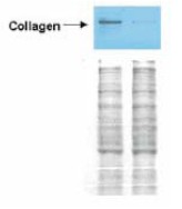
Western blot analysis is shown using Collagen type I antibody to detect expression of collagen I in Wistar rat hepatic stellate cells (HSC) in control (GFP-transduced) (left lane) and PPARgamma-transduced cell lysates (right lane). Protein staining shown below each blot depicts equal protein loading. An equal amount of the whole cell protein (100 ug) was separated by SDS-PAGE and electroblotted to nitrocellulose membranes. Proteins were detected by incubating the membrane with Collagen type I antibody at a concentration of 0.2-2 ug/10 ml in TBS (100 mM Tris-HCl, 0.15 M NaCl, pH 7.4) with 5% Non-fat milk. Detection occurred by incubation with a horseradish peroxidase-conjugated secondary antibody at 1 ug/10 ml. Proteins were detected by a chemiluminescent method using the PIERCE ECL kit (Amersham Biosciences). Other detection systems will yield similar results. See Hazra et al. (2004) for additional details.
Western blot analysis is shown using Collagen type I antibody to detect expression of collagen I in Wistar rat hepatic stellate cells (HSC) in control (GFP-transduced) (left lane) and PPARgamma-transduced cell lysates (right lane). Protein staining shown below each blot depicts equal protein loading. An equal amount of the whole cell protein (100 ug) was separated by SDS-PAGE and electroblotted to nitrocellulose membranes. Proteins were detected by incubating the membrane with Collagen type I antibody at a concentration of 0.2-2 ug/10 ml in TBS (100 mM Tris-HCl, 0.15 M NaCl, pH 7.4) with 5% Non-fat milk. Detection occurred by incubation with a horseradish peroxidase-conjugated secondary antibody at 1 ug/10 ml. Proteins were detected by a chemiluminescent method using the PIERCE ECL kit (Amersham Biosciences). Other detection systems will yield similar results. See Hazra et al. (2004) for additional details.
Collagen I (COL1A1) Rabbit Polyclonal Antibody
R1038
ApplicationsImmunoPrecipitation, Western Blot, ELISA, ImmunoHistoChemistry
Product group Antibodies
ReactivityBovine, Human, Mammals, Mouse, Rat
TargetCOL1A1
Overview
- SupplierOriGene
- Product NameCollagen I (COL1A1) Rabbit Polyclonal Antibody
- Delivery Days Customer14
- ApplicationsImmunoPrecipitation, Western Blot, ELISA, ImmunoHistoChemistry
- CertificationResearch Use Only
- ClonalityPolyclonal
- Gene ID1277
- Target nameCOL1A1
- Target descriptioncollagen type I alpha 1 chain
- Target synonymsalpha-1 type I collagen; alpha1(I) procollagen; CAFYD; collagen alpha 1 chain type I; collagen alpha-1(I) chain; collagen alpha-1(I) chain preproprotein; collagen of skin, tendon and bone, alpha-1 chain; collagen, type I, alpha 1; EDSARTH1; EDSC; OI1; OI2; OI3; OI4; pro-alpha-1 collagen type 1; type I proalpha 1; type I procollagen alpha 1 chain
- HostRabbit
- Protein IDP02452
- Protein NameCollagen alpha-1(I) chain
- Scientific DescriptionCollagen I (COL1A1) rabbit polyclonal antibody, Purified
- ReactivityBovine, Human, Mammals, Mouse, Rat
- Storage Instruction-20°C,2°C to 8°C
- UNSPSC12352203