

Connexin 43 antibody [CXN-6]
GTX11369
ApplicationsImmunoFluorescence, Western Blot, ELISA, ImmunoCytoChemistry, ImmunoHistoChemistry, ImmunoHistoChemistry Frozen, ImmunoHistoChemistry Paraffin
Product group Antibodies
ReactivityBovine, Hamster, Human, Mouse, Rat
TargetGJA1
Overview
- SupplierGeneTex
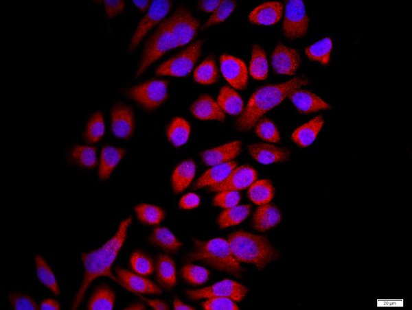
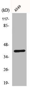
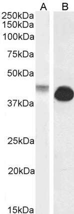
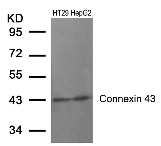
- Product NameConnexin 43 antibody [CXN-6]
- Delivery Days Customer9
- Application Supplier NoteWB: 1:8,000-1:16.000. *Optimal dilutions/concentrations should be determined by the researcher.Not tested in other applications.
- ApplicationsImmunoFluorescence, Western Blot, ELISA, ImmunoCytoChemistry, ImmunoHistoChemistry, ImmunoHistoChemistry Frozen, ImmunoHistoChemistry Paraffin
- CertificationResearch Use Only
- ClonalityMonoclonal
- Clone IDCXN-6
- ConjugateUnconjugated
- Gene ID2697
- Target nameGJA1
- Target descriptiongap junction protein alpha 1
- Target synonymsAVSD3, CMDR, CX43, EKVP, EKVP3, GJAL, HLHS1, HSS, ODDD, PPKCA, gap junction alpha-1 protein, connexin-43, gap junction 43 kDa heart protein, gap junction protein, alpha 1, 43kDa
- HostMouse
- IsotypeIgM
- Protein IDP17302
- Protein NameGap junction alpha-1 protein
- Scientific DescriptionGap junctions are aggregations of intercellular channels, which directly connect the cytoplasm of adjacent cells. Gap junctions coordinate cellular and organ function in tissues and are involved in metabolic cooperation between cells, synchronization of cellular physiological activities, growth control, and developmental regulation. The gap junction channels allow intercellular exchange of ions, nucleotides, and small molecules between adjacent cells. Unlike other membrane channels, intercellular channels span two plasma membranes and require the contribution of hemi-channels, called connexons, from both participating cells. These channels accommodate molecules as large as 1 kDa. They have been detected in virtually every cell type in mammals, except mature skeletal muscle, spermatozoa, and erythrocytes. Two connexons interact in the extracellular space to form the complete intercellular channel. Each connexon is composed of six similar or identical proteins, which have been termed connexins. Connexins (Cx) are a multi-gene family of highly related proteins with molecular weights between 26 and 70 kDa. At least a dozen distinct connexin genes have been identified; many of them expressed in a tissue-specific manner. Two distinct lineages have been identified in mammals. One termed class I or beta group consists of Cx26, Cx30, Cx31, Cx31.1, and Cx32. The other, termed class II or alpha group, is represented by Cx33, Cx37, Cx40, Cx43 and Cx46. All of the connexins share a common membrane topology but differ in their conductance and channel gating properties. The structure of connexin molecules includes a cytoplasmic N-terminal region, four transmembrane domains, two extracellular loops, and a C-terminal cytoplasmic tail of varying length. The various connexins are highly conserved in the transmembrane and extracellular regions, but they differ in their cytoplasmic domain. The 43 kDa connexin protein (connexin-43, Cx43) belongs to the alpha type (group II) subfamily of connexin proteins. It is expressed in most tissues, even though the pattern of expression may differ in various cell types. For example, in the brain it is found in astrocytes, ependyma and leptomeninges but not in neurons, oligodendrocytes and pinealocytes; in the liver it is present in Ito cells but not hepatocytes. Gap junction protein levels change in response to disruption of tissue architecture. For instance, an increased expression of Cx43 was found in early stages of atherosclerosis. Changes in the levels or types of connexin expressed in a given cell type have been found to correlate with tumor progression and metastasis. However, glioma cells transfected with the oncogene neu (c-erb-B2) have been shown to exhibit a major reduction in intercellular communication with no decrease in overall expression of Cx43. Monoclonal antibodies reacting specifically with Cx43 may be applied in diverse cellular and molecular approaches to study gap junctions and their properties. They can also be used to correlate their expression pattern with physiological functions or pathological conditions.
- ReactivityBovine, Hamster, Human, Mouse, Rat
- Storage Instruction-20°C or -80°C,2°C to 8°C
- UNSPSC12352203
References
- Lao S, Xu J, Liu Y, et al. A comparative study of the influence of two types of PHEMA stents on the differentiation of ASCs to myocardial cells. Mol Med Rep. 2017,16(1):507-514. doi: 10.3892/mmr.2017.6680Read this paper
Datasheet
Related products

Product group Antibodies
Anti-Connexin 43/GJA1 Antibody Picoband(r)A00599-CARRIER-FREE
ApplicationsImmunoFluorescence, Western Blot, ELISA, ImmunoCytoChemistry, ImmunoHistoChemistry
ReactivityHuman, Mouse, Rat
TargetGJA1
- SizePrice
Product group Antibodies
Anti-GJA1 Antibody144-02163
ApplicationsWestern Blot
ReactivityHuman, Mouse
TargetGJA1
- SizePrice
Product group Antibodies
ApplicationsImmunoPrecipitation, Western Blot, ImmunoCytoChemistry, ImmunoHistoChemistry
ReactivityMouse
TargetGJA1
- SizePrice
Product group Antibodies
References
Connexin 43 Polyclonal AntibodyBS-0651R
ApplicationsFlow Cytometry, ImmunoFluorescence, Western Blot, ELISA, ImmunoCytoChemistry, ImmunoHistoChemistry, ImmunoHistoChemistry Frozen, ImmunoHistoChemistry Paraffin
ReactivityBovine, Canine, Chicken, Human, Mouse, Rat
TargetGJA1
- SizePrice
Product group Antibodies
GJA1 AntibodyCSB-PA001755
ApplicationsImmunoFluorescence, Western Blot, ELISA, ImmunoHistoChemistry
ReactivityHuman, Mouse, Rat
TargetGJA1
- SizePrice
Product group Antibodies
ApplicationsWestern Blot, ELISA
ReactivityHuman, Rat
- SizePrice

Product group Antibodies
Connexin 43 antibody, InternalGTX88424
ApplicationsWestern Blot
ReactivityHuman, Rat
TargetGJA1
- SizePrice
Product group Antibodies
References
Connexin 43 antibodyGTX50571
ApplicationsWestern Blot, ImmunoHistoChemistry, ImmunoHistoChemistry Paraffin
ReactivityHuman, Rabbit
TargetGJA1
- SizePrice