

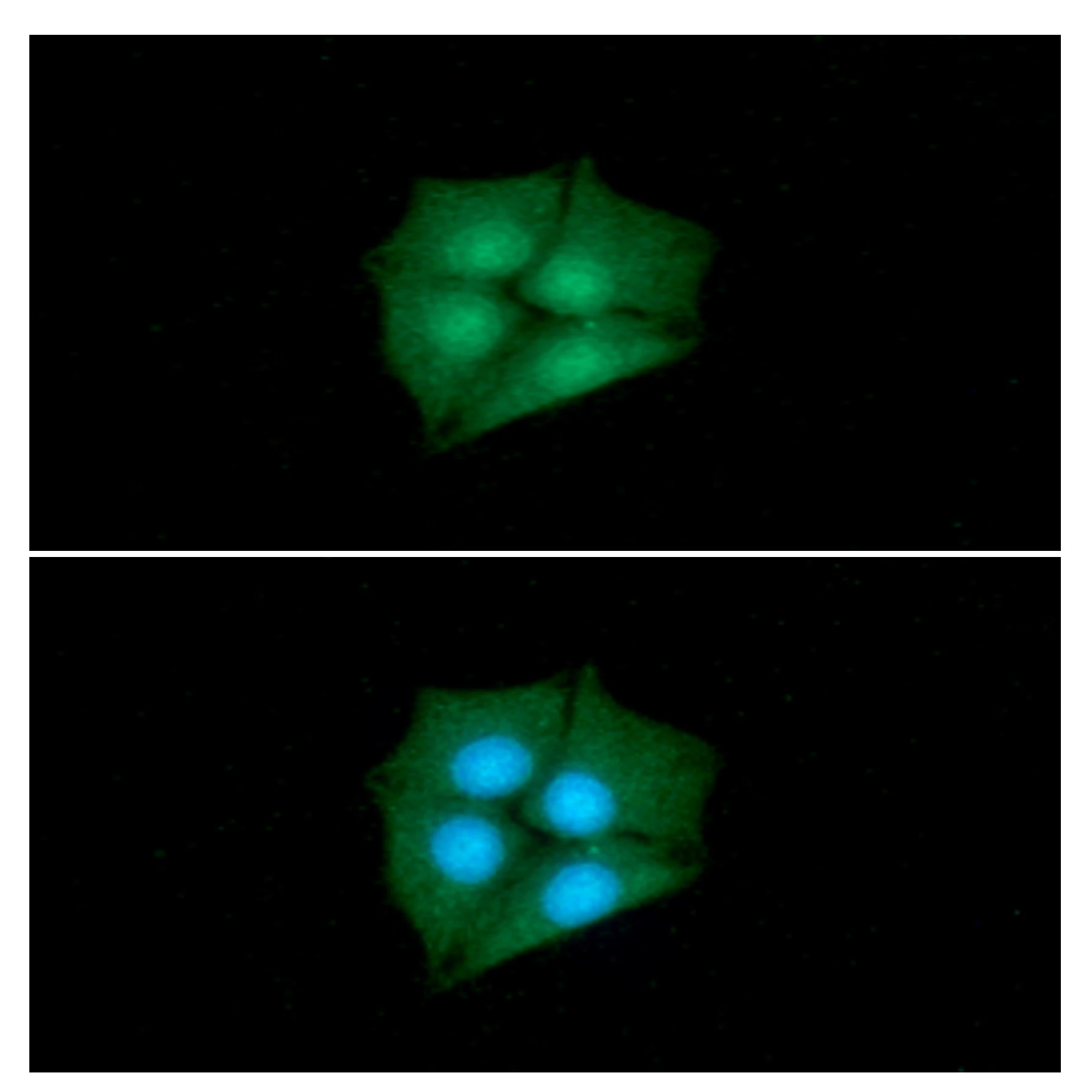
ICC/IF analysis of 3T3 cells using GTX50013 CRABP1 antibody [AT1A1]. Dilution : 1:100
![WB analysis of mouse eye tissue lysate using GTX50013 CRABP1 antibody [AT1A1]. Loading : 30 μg Dilution : 1:500](https://www.genetex.com/upload/website/prouct_img/normal/GTX50013/GTX50013_20231002_WB_23100123_253.webp "WB analysis of mouse eye tissue lysate using GTX50013 CRABP1 antibody [AT1A1]. Loading : 30 μg Dilution : 1:500")
![FACS analysis of 3T3 cells using GTX50013 CRABP1 antibody [AT1A1]. Red : Primary antibody Black : Isotype control](https://www.genetex.com/upload/website/prouct_img/normal/GTX50013/GTX50013_20231002_FACS_23100123_142.webp "FACS analysis of 3T3 cells using GTX50013 CRABP1 antibody [AT1A1]. Red : Primary antibody Black : Isotype control")
![WB analysis of cell lysate using GTX50013 CRABP1 antibody [AT1A1]. Lane 1 : MCF-7 Loading : 40 μg Dilution : 1:500](https://www.genetex.com/upload/website/prouct_img/normal/GTX50013/GTX50013_20231002_WB_1_23100123_383.webp "WB analysis of cell lysate using GTX50013 CRABP1 antibody [AT1A1]. Lane 1 : MCF-7 Loading : 40 μg Dilution : 1:500")
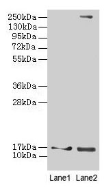
![WB analysis of cell lysates using GTX50013 CRABP1 antibody [AT1A1]. Lane 1 : 293T cell lysate Lane 2 : CRABP1 Transfected 293T cell lysate Loading : 20 μg Dilution : 1:500](https://www.genetex.com/upload/website/prouct_img/normal/GTX50013/GTX50013_20231002_WB_2_23100123_474.webp "WB analysis of cell lysates using GTX50013 CRABP1 antibody [AT1A1]. Lane 1 : 293T cell lysate Lane 2 : CRABP1 Transfected 293T cell lysate Loading : 20 μg Dilution : 1:500")
ICC/IF analysis of 3T3 cells using GTX50013 CRABP1 antibody [AT1A1]. Dilution : 1:100
CRABP1 antibody [AT1A1]
GTX50013
ApplicationsFlow Cytometry, ImmunoFluorescence, Western Blot, ELISA, ImmunoCytoChemistry
Product group Antibodies
ReactivityHuman, Mouse
TargetCRABP1
Overview
- SupplierGeneTex
- Product NameCRABP1 antibody [AT1A1]
- Delivery Days Customer9
- Application Supplier NoteWe recommend the following starting dilutions:Western Blot: Use at 1:500 ~ 1000.Optimal working concentrations should be determined experimentally by the end user.
- ApplicationsFlow Cytometry, ImmunoFluorescence, Western Blot, ELISA, ImmunoCytoChemistry
- CertificationResearch Use Only
- ClonalityMonoclonal
- Clone IDAT1A1
- Concentration1 mg/ml
- ConjugateUnconjugated
- Gene ID1381
- Target nameCRABP1
- Target descriptioncellular retinoic acid binding protein 1
- Target synonymsCRABP, CRABP-I, CRABPI, RBP5, cellular retinoic acid-binding protein 1, cellular retinoic acid-binding protein I
- HostMouse
- IsotypeIgG2b
- Protein IDP29762
- Protein NameCellular retinoic acid-binding protein 1
- Scientific DescriptionThis gene encodes a specific binding protein for a vitamin A family member and is thought to play an important role in retinoic acid-mediated differentiation and proliferation processes. It is structurally similar to the cellular retinol-binding proteins, but binds only retinoic acid at specific sites within the nucleus, which may contribute to vitamin A-directed differentiation in epithelial tissue. [provided by RefSeq, Jul 2008]
- ReactivityHuman, Mouse
- Storage Instruction-20°C or -80°C,2°C to 8°C
- UNSPSC41116161
Datasheet
Related products
Product group Antibodies
Anti-CRABP1 (C-term) Antibody102-26836
ApplicationsWestern Blot, ImmunoHistoChemistry, ImmunoHistoChemistry Paraffin
TargetCRABP1
- SizePrice

Product group Antibodies
Anti-CRABP1 Antibody Picoband(r)A07951-1-CARRIER-FREE
ApplicationsFlow Cytometry, Western Blot, ELISA
ReactivityHuman
TargetCRABP1
- SizePrice
Product group Antibodies

CRABP1 / CRABP AntibodyLS-C830465
ApplicationsWestern Blot, ELISA, ImmunoHistoChemistry
ReactivityHuman, Mouse, Rat
TargetCRABP1
- SizePrice
Product group Antibodies
CRABP1 Recombinant AntibodyBSM-62149R
ApplicationsImmunoPrecipitation, Western Blot
ReactivityHuman, Mouse
TargetCRABP1
- SizePrice
Product group Antibodies
Crabp1 Polyclonal AntibodyCAC08331
ApplicationsWestern Blot, ELISA, ImmunoHistoChemistry
ReactivityMouse
TargetCRABP1
- SizePrice
Product group Antibodies
CRABP1 AntibodyCSB-PA005935DA01HU
ApplicationsWestern Blot, ELISA, ImmunoHistoChemistry
ReactivityHuman, Mouse
TargetCRABP1
- SizePrice

Product group Antibodies
Anti-CRABP1 AntibodyHPA017203
ApplicationsImmunoHistoChemistry
ReactivityHuman
TargetCRABP1
- SizePrice
Product group Antibodies
ApplicationsImmunoFluorescence, Western Blot, ELISA, ImmunoCytoChemistry
ReactivityHuman
TargetCRABP1
- SizePrice