

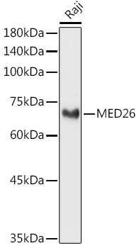
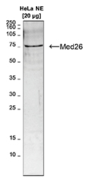
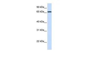
Western blot analysis of various lysates using MED26 Rabbit pAb (TA378438) at 1:1000 dilution.
Secondary antibody: HRP-conjugated Goat anti-Rabbit IgG (H+L) at 1:10000 dilution.
Lysates/proteins: 25μg per lane.
Blocking buffer: 3% nonfat dry milk in TBST.
Detection: ECL Basic Kit .
Exposure time: 180s.
Secondary antibody: HRP-conjugated Goat anti-Rabbit IgG (H+L) at 1:10000 dilution.
Lysates/proteins: 25μg per lane.
Blocking buffer: 3% nonfat dry milk in TBST.
Detection: ECL Basic Kit .
Exposure time: 180s.
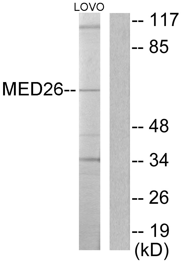
CRSP7 (MED26) Rabbit Polyclonal Antibody
TA378438
ApplicationsWestern Blot
Product group Antibodies
ReactivityHuman, Mouse, Rat
TargetMED26
Overview
- SupplierOriGene
- Product NameCRSP7 (MED26) Rabbit Polyclonal Antibody
- Delivery Days Customer14
- ApplicationsWestern Blot
- CertificationResearch Use Only
- ClonalityPolyclonal
- Gene ID9441
- Target nameMED26
- Target descriptionmediator complex subunit 26
- Target synonymsCRSP7, CRSP70, mediator of RNA polymerase II transcription subunit 26, ARC70, CRSP complex subunit 7, activator-recruited cofactor 70 kDa component, cofactor required for Sp1 transcriptional activation subunit 7, cofactor required for Sp1 transcriptional activation, subunit 7 (70kD), cofactor required for Sp1 transcriptional activation, subunit 7, 70kDa, transcriptional coactivator CRSP70
- HostRabbit
- IsotypeIgG
- Protein IDO95402
- Protein NameMediator of RNA polymerase II transcription subunit 26
- Scientific DescriptionMED26 Rabbit polyclonal Antibody
- ReactivityHuman, Mouse, Rat
- Storage Instruction-20°C
- UNSPSC12352203
MSDS
Related products
Product group Antibodies
Anti-MED26 AntibodyA99042
ApplicationsWestern Blot, ELISA
ReactivityHuman, Mouse
- SizePrice
Product group Antibodies
Anti-MED26 (Center) Antibody102-22299
ApplicationsWestern Blot
TargetMED26
- SizePrice

Product group Antibodies
Anti-MED26 Antibody Picoband(r)A09340-1-CARRIER-FREE
ApplicationsFlow Cytometry, ImmunoFluorescence, Western Blot, ELISA, ImmunoCytoChemistry, ImmunoHistoChemistry
ReactivityHuman
TargetMED26
- SizePrice
Product group Antibodies
Med26 Polyclonal AntibodyBS-53124R
ApplicationsWestern Blot
ReactivityHuman
TargetMED26
- SizePrice
Product group Antibodies
MED26 AntibodyCSB-PA013659LA01HU
ApplicationsELISA, ImmunoHistoChemistry
ReactivityHuman
TargetMED26
- SizePrice
Product group Antibodies

MED26 / CRSP7 Antibody (aa543-593)LS-C289120
ApplicationsImmunoPrecipitation
ReactivityHuman
TargetMED26
- SizePrice

Product group Antibodies
Anti-MED26 AntibodyHPA071835
ApplicationsChIP Chromatin ImmunoPrecipitation, ImmunoCytoChemistry
ReactivityHuman
TargetMED26
- SizePrice
Product group Antibodies
MED26 antibody, N-termGTX49066
ApplicationsWestern Blot
ReactivityHuman
TargetMED26
- SizePrice