

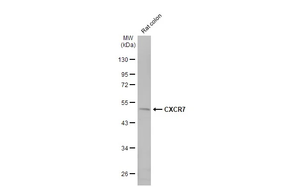
Rat tissue extract (50 μg) was separated by 10% SDS-PAGE, and the membrane was blotted with CXCR7 antibody [HL2189] (GTX638193) diluted at 1:1000. The HRP-conjugated anti-rabbit IgG antibody (GTX213110-01) was used to detect the primary antibody.
![Mouse tissue extract (50 μg) was separated by 10% SDS-PAGE, and the membrane was blotted with CXCR7 antibody [HL2189] (GTX638193) diluted at 1:1000. The HRP-conjugated anti-rabbit IgG antibody (GTX213110-01) was used to detect the primary antibody.](https://www.genetex.com/upload/website/prouct_img/normal/GTX638193/GTX638193_T-44942_20230707_WB_M_spleen_23071223_941.webp "Mouse tissue extract (50 μg) was separated by 10% SDS-PAGE, and the membrane was blotted with CXCR7 antibody [HL2189] (GTX638193) diluted at 1:1000. The HRP-conjugated anti-rabbit IgG antibody (GTX213110-01) was used to detect the primary antibody.")
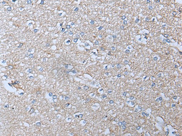
![CXCR7 antibody [HL2189] detects CXCR7 protein at cell membrane by immunohistochemical analysis. Sample: Paraffin-embedded human colon cancer. CXCR7 stained by CXCR7 antibody [HL2189] (GTX638193) diluted at 1:100. Antigen Retrieval: Citrate buffer, pH 6.0, 15 min](https://www.genetex.com/upload/website/prouct_img/normal/GTX638193/GTX638193_T-44942_20230728_IHC-P_1_23080901_231.webp "CXCR7 antibody [HL2189] detects CXCR7 protein at cell membrane by immunohistochemical analysis. Sample: Paraffin-embedded human colon cancer. CXCR7 stained by CXCR7 antibody [HL2189] (GTX638193) diluted at 1:100. Antigen Retrieval: Citrate buffer, pH 6.0, 15 min")
![CXCR7 antibody [HL2189] detects CXCR7 protein at cell membrane by immunohistochemical analysis. Sample: Paraffin-embedded Raji xenograft. CXCR7 stained by CXCR7 antibody [HL2189] (GTX638193) diluted at 1:100. Antigen Retrieval: Citrate buffer, pH 6.0, 15 min](https://www.genetex.com/upload/website/prouct_img/normal/GTX638193/GTX638193_T-44942_20230728_IHC-P_23080901_299.webp "CXCR7 antibody [HL2189] detects CXCR7 protein at cell membrane by immunohistochemical analysis. Sample: Paraffin-embedded Raji xenograft. CXCR7 stained by CXCR7 antibody [HL2189] (GTX638193) diluted at 1:100. Antigen Retrieval: Citrate buffer, pH 6.0, 15 min")
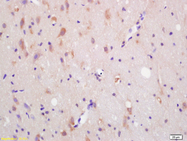
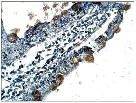
![CXCR7 antibody [HL2189] detects CXCR7 protein at cell membrane by immunohistochemical analysis. Sample: Paraffin-embedded rat colon. CXCR7 stained by CXCR7 antibody [HL2189] (GTX638193) diluted at 1:100. Antigen Retrieval: Citrate buffer, pH 6.0, 15 min](https://www.genetex.com/upload/website/prouct_img/normal/GTX638193/GTX638193_T-44942_20230728_IHC-P_R_23080901_998.webp "CXCR7 antibody [HL2189] detects CXCR7 protein at cell membrane by immunohistochemical analysis. Sample: Paraffin-embedded rat colon. CXCR7 stained by CXCR7 antibody [HL2189] (GTX638193) diluted at 1:100. Antigen Retrieval: Citrate buffer, pH 6.0, 15 min")
![Unboiled Raji whole cell and membrane extracts (30 μg) were separated by 10% SDS-PAGE, and the membrane was blotted with CXCR7 antibody [HL2189] (GTX638193) diluted at 1:50000. The HRP-conjugated anti-rabbit IgG antibody (GTX213110-01) was used to detect the primary antibody. (WCE: whole cell extract; ME: membrane extract)](https://www.genetex.com/upload/website/prouct_img/normal/GTX638193/GTX638193_45159_20230908_WB_Fraction_23091319_457.webp "Unboiled Raji whole cell and membrane extracts (30 μg) were separated by 10% SDS-PAGE, and the membrane was blotted with CXCR7 antibody [HL2189] (GTX638193) diluted at 1:50000. The HRP-conjugated anti-rabbit IgG antibody (GTX213110-01) was used to detect the primary antibody. (WCE: whole cell extract; ME: membrane extract)")
![Non-transfected (–) and transfected (+) SW480 whole cell extracts (30 μg) were separated by 10% SDS-PAGE, and the membrane was blotted with CXCR7 antibody [HL2189] (GTX638193) diluted at 1:10000. The HRP-conjugated anti-rabbit IgG antibody (GTX213110-01) was used to detect the primary antibody.](https://www.genetex.com/upload/website/prouct_img/normal/GTX638193/GTX638193_45159_20230915_WB_shRNA_watermark_23091901_973.webp "Non-transfected (–) and transfected (+) SW480 whole cell extracts (30 μg) were separated by 10% SDS-PAGE, and the membrane was blotted with CXCR7 antibody [HL2189] (GTX638193) diluted at 1:10000. The HRP-conjugated anti-rabbit IgG antibody (GTX213110-01) was used to detect the primary antibody.")
![CXCR7 antibody [HL2189] detects CXCR7 protein by immunofluorescent analysis. Sample: Mock and transfected 293T cells were fixed in 4% paraformaldehyde at RT for 15 min. Green: CXCR7 stained by CXCR7 antibody [HL2189] (GTX638193) diluted at 1:500. Blue: Fluoroshield with DAPI (GTX30920). Scale bar= 10μm.](https://www.genetex.com/upload/website/prouct_img/normal/GTX638193/GTX638193_45159_20231110_ICC_IF_B_23111422_223.webp "CXCR7 antibody [HL2189] detects CXCR7 protein by immunofluorescent analysis. Sample: Mock and transfected 293T cells were fixed in 4% paraformaldehyde at RT for 15 min. Green: CXCR7 stained by CXCR7 antibody [HL2189] (GTX638193) diluted at 1:500. Blue: Fluoroshield with DAPI (GTX30920). Scale bar= 10μm.")
![CXCR7 antibody [HL2189] detects CXCR7 protein at cell membrane by immunohistochemical analysis. Sample: Paraffin-embedded Raji xenograft. CXCR7 stained by CXCR7 antibody [HL2189] (GTX638193) diluted at 1:200 and competitor's antibody (MAB42273) diluted at 1:200. Antigen Retrieval: Citrate buffer, pH 6.0, 15 min *Competitor's antibody is not affiliated with GeneTex and does not endorse this product.](https://www.genetex.com/upload/website/prouct_img/normal/GTX638193/GTX638193_45159_20240301_IHC-P_competitor_24030600_600.webp "CXCR7 antibody [HL2189] detects CXCR7 protein at cell membrane by immunohistochemical analysis. Sample: Paraffin-embedded Raji xenograft. CXCR7 stained by CXCR7 antibody [HL2189] (GTX638193) diluted at 1:200 and competitor's antibody (MAB42273) diluted at 1:200. Antigen Retrieval: Citrate buffer, pH 6.0, 15 min *Competitor's antibody is not affiliated with GeneTex and does not endorse this product.")
![CXCR7 antibody [HL2189] detects CXCR7 protein by immunofluorescent analysis. Sample: THP-1 cells were fixed in 4% paraformaldehyde at RT for 15 min. Green: CXCR7 stained by CXCR7 antibody [HL2189] (GTX638193) diluted at 1:100. Blue: Fluoroshield with DAPI (GTX30920).](https://www.genetex.com/upload/website/prouct_img/normal/GTX638193/GTX638193_45159_20241122_ICC_IF_24120522_412.webp "CXCR7 antibody [HL2189] detects CXCR7 protein by immunofluorescent analysis. Sample: THP-1 cells were fixed in 4% paraformaldehyde at RT for 15 min. Green: CXCR7 stained by CXCR7 antibody [HL2189] (GTX638193) diluted at 1:100. Blue: Fluoroshield with DAPI (GTX30920).")
Rat tissue extract (50 μg) was separated by 10% SDS-PAGE, and the membrane was blotted with CXCR7 antibody [HL2189] (GTX638193) diluted at 1:1000. The HRP-conjugated anti-rabbit IgG antibody (GTX213110-01) was used to detect the primary antibody.
CXCR7 antibody [HL2189]
GTX638193
ApplicationsImmunoFluorescence, Western Blot, ImmunoCytoChemistry, ImmunoHistoChemistry, ImmunoHistoChemistry Frozen, ImmunoHistoChemistry Paraffin
Product group Antibodies
ReactivityHuman, Mouse, Rat
TargetACKR3
Overview
- SupplierGeneTex
- Product NameCXCR7 antibody [HL2189]
- Delivery Days Customer9
- Application Supplier NoteWB: 1:500-1:3000. *Optimal dilutions/concentrations should be determined by the researcher.Not tested in other applications.
- ApplicationsImmunoFluorescence, Western Blot, ImmunoCytoChemistry, ImmunoHistoChemistry, ImmunoHistoChemistry Frozen, ImmunoHistoChemistry Paraffin
- CertificationResearch Use Only
- ClonalityMonoclonal
- Clone IDHL2189
- Concentration1 mg/ml
- ConjugateUnconjugated
- Gene ID57007
- Target nameACKR3
- Target descriptionatypical chemokine receptor 3
- Target synonymsCMKOR1, CXC-R7, CXCR-7, CXCR7, GPR159, RDC-1, RDC1, atypical chemokine receptor 3, C-X-C chemokine receptor type 7, G protein-coupled receptor, G-protein coupled receptor 159, G-protein coupled receptor RDC1 homolog, chemokine (C-X-C motif) receptor 7, chemokine orphan receptor 1
- HostRabbit
- IsotypeIgG
- Protein IDP25106
- Protein NameAtypical chemokine receptor 3
- Scientific DescriptionThis gene encodes a member of the G-protein coupled receptor family. Although this protein was earlier thought to be a receptor for vasoactive intestinal peptide (VIP), it is now considered to be an orphan receptor, in that its endogenous ligand has not been identified. The protein is also a coreceptor for human immunodeficiency viruses (HIV). Translocations involving this gene and HMGA2 on chromosome 12 have been observed in lipomas. [provided by RefSeq, Jul 2008]
- ReactivityHuman, Mouse, Rat
- Storage Instruction-20°C or -80°C,2°C to 8°C
- UNSPSC41116161
Datasheet
Related products
Product group Antibodies
Anti-ACKR3 AntibodyA46576
ApplicationsImmunoHistoChemistry
ReactivityHuman
- SizePrice

Product group Antibodies
Anti-GPCR RDC1/CXCR-7/ACKR3 Antibody Picoband(r)A02656-2-CARRIER-FREE
ApplicationsFlow Cytometry, Western Blot, ImmunoHistoChemistry
ReactivityHuman
TargetACKR3
- SizePrice
Product group Antibodies
Anti-ACKR3 Antibody144-12712
ApplicationsWestern Blot
ReactivityHuman, Mouse, Rat
TargetACKR3
- SizePrice
Product group Antibodies

ACKR3 / CXCR7 AntibodyLS-C747794
ApplicationsWestern Blot
ReactivityHuman, Mouse, Rat
TargetACKR3
- SizePrice
Product group Antibodies
References
ACKR3 Polyclonal AntibodyBS-4897R
ApplicationsImmunoFluorescence, Western Blot, ELISA, ImmunoCytoChemistry, ImmunoHistoChemistry, ImmunoHistoChemistry Frozen, ImmunoHistoChemistry Paraffin
ReactivityBovine, Canine, Equine, Human, Mouse, Porcine, Rabbit, Rat
TargetACKR3
- SizePrice
Product group Antibodies
ACKR3 AntibodyCSB-PA001850
ApplicationsImmunoFluorescence, Western Blot, ELISA
ReactivityHuman, Monkey, Mouse, Rat
TargetACKR3
- SizePrice
Product group Antibodies

Goat anti-CXCR7 / RDC1EB12777
ApplicationsWestern Blot, ELISA
ReactivityHuman
TargetACKR3
- SizePrice
![Boiled and unboiled Raji whole cell and membrane extracts (30 μg) were separated by 10% SDS-PAGE, and the membrane was blotted with CXCR7 antibody [C1C2], Internal (GTX100027) diluted at 1:5000. The HRP-conjugated anti-rabbit IgG antibody (GTX213110-01) was used to detect the primary antibody, and the signal was developed with Trident ECL plus-Enhanced. (WCE: whole cell extract; ME: membrane extract)](https://www.genetex.com/upload/website/prouct_img/normal/GTX100027/GTX100027_39210_20230505_WB_Fraction_23050918_504.webp)
Product group Antibodies
CXCR7 antibody [C1C2], InternalGTX100027
ApplicationsFlow Cytometry, ImmunoFluorescence, ImmunoPrecipitation, Western Blot, ELISA, ImmunoCytoChemistry, ImmunoHistoChemistry, ImmunoHistoChemistry Frozen, ImmunoHistoChemistry Paraffin
ReactivityHuman, Mouse, Rat
TargetACKR3
- SizePrice
Product group Antibodies
References
CXCR7 antibodyGTX82935
ApplicationsImmunoPrecipitation, Western Blot, ELISA, ImmunoHistoChemistry, ImmunoHistoChemistry Paraffin
ReactivityHuman, Monkey, Mouse, Primate, Rat
TargetACKR3
- SizePrice
![IHC-P analysis of human glioblastoma (GBM) tissue using GTX641403 CXCR7 antibody [H342] HistoMAX?. Glioblastoma with strong CXCR7 staining of most tumor cells.](https://www.genetex.com/upload/website/prouct_img/normal/GTX641403/GTX641403_20250117_IHC-P_1_25012200_702.webp)
Product group Antibodies
CXCR7 antibody [H342] HistoMAX(tm)GTX641403
ApplicationsImmunoHistoChemistry, ImmunoHistoChemistry Paraffin
ReactivityHuman
TargetACKR3
- SizePrice