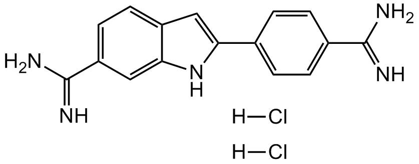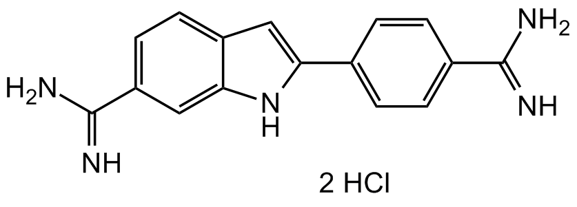
Chemical Structure
DAPI . dihydrochloride [28718-90-3] [28718-90-3]
AG-CR1-3668
CAS Number28718-90-3
Product group Chemicals
Estimated Purity>97%
Molecular Weight277.3 . 72.9
Overview
- SupplierAdipoGen Life Sciences
- Product NameDAPI . dihydrochloride [28718-90-3] [28718-90-3]
- Delivery Days Customer10
- CAS Number28718-90-3
- CertificationResearch Use Only
- Estimated Purity>97%
- Hazard InformationWarning
- Molecular FormulaC16H15N5 . 2HCl
- Molecular Weight277.3 . 72.9
- Scientific DescriptionCell permeable fluorescent DNA stain. AT-sequence-specific DNA intercalator. Binds to the minor groove of double-stranded DNA (preferentially to adenine and thymine (AT) rich DNA), like Hoechst dyes, forming a stable complex which fluoresces approximately 20 times greater than DAPI alone. Spectral Data: Excitation: lambdaex 340 nm; Emission: lambdaem 488 nm (only DAPI). Excitation: lambdaex 360nm; Emission: lambdaem 460nm (DAPI-DNA complex). Commonly used to stain DNA and chromosomes for fluorescent microscopy and flow cytometry applications. DAPI is often used as a counterstain, as its ultraviolet excitation (lambdaex 360nm) and blue emission (lambdaem 460nm) wavelengths separate it nicely from many popular primary fluorophores. Can be used on either fixed or live cells, although it passes through the membrane less efficiently in live cells and therefore the effectiveness of the stain is lower and demands higher concentrations to be used. Reversible inhibitor of S-adenosyl-L-methionine decarboxylase and KAO (diamine oxidase). - Chemical. CAS: 28718-90-3. Formula: C16H15N5 . 2HCl. MW: 277.3 . 72.9. Synthetic. Cell permeable fluorescent DNA stain. AT-sequence-specific DNA intercalator. Binds to the minor groove of double-stranded DNA (preferentially to adenine and thymine (AT) rich DNA), like Hoechst dyes, forming a stable complex which fluoresces approximately 20 times greater than DAPI alone. Spectral Data: Excitation: lambdaex 340 nm; Emission: lambdaem 488 nm (only DAPI). Excitation: lambdaex 360nm; Emission: lambdaem 460nm (DAPI-DNA complex). Commonly used to stain DNA and chromosomes for fluorescent microscopy and flow cytometry applications. DAPI is often used as a counterstain, as its ultraviolet excitation (lambdaex 360nm) and blue emission (lambdaem 460nm) wavelengths separate it nicely from many popular primary fluorophores. Can be used on either fixed or live cells, although it passes through the membrane less efficiently in live cells and therefore the effectiveness of the stain is lower and demands higher concentrations to be used. Reversible inhibitor of S-adenosyl-L-methionine decarboxylase and KAO (diamine oxidase).
- SMILES[H]Cl.[H]Cl.NC(C1=CC=C2C(NC(C3=CC=C(C(N)=N)C=C3)=C2)=C1)=N
- Storage Instruction-20°C,2°C to 8°C
- UNSPSC12162000

![DAPI [28718-90-3]](https://www.antibodies.com/image/catalog/319/A319637_1.jpg)


![DAPI Dihydrochloride [28718-90-3]](https://www.targetmol.com/group3/M00/35/78/CgoaEGayIC-EW36wAAAAAPsOgkI503.png)