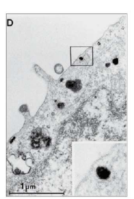
Figure 1. Electron Microscopy Image of internalized EGF-R antibody complexes in A431 cells. The cells were stained with AM20237PU-N/AM20237PU-S for 1h at 4°C, followd by Peroxidase-conjugated Goat anti-Mouse IgG. After fixation with 2.5% glutaraldehyde, the signal was detected using DAB (0.6 mg/ml) for 20 min at RT. The cells were then processed for the preparation of ultrathin sections. Reins HA at al. (1993) J. Cell. Biochem 51 (2): 236-48.
EGFR (Extracell. non Ligand binding Site) Mouse Monoclonal Antibody [Clone ID: EGF-R2]

AM20237PU-S
ApplicationsFlow Cytometry, ELISA, ImmunoHistoChemistry
Product group Antibodies
ReactivityHuman
TargetEGFR
Overview
- SupplierOriGene
- Product NameEGFR (Extracell. non Ligand binding Site) Mouse Monoclonal Antibody [Clone ID: EGF-R2]
- Delivery Days Customer14
- ApplicationsFlow Cytometry, ELISA, ImmunoHistoChemistry
- CertificationResearch Use Only
- ClonalityMonoclonal
- Clone IDEGF-R2
- Gene ID1956
- Target nameEGFR
- Target descriptionepidermal growth factor receptor
- Target synonymsavian erythroblastic leukemia viral (v-erb-b) oncogene homolog; cell growth inhibiting protein 40; cell proliferation-inducing protein 61; epidermal growth factor receptor; epidermal growth factor receptor tyrosine kinase domain; ERBB; ERBB1; erb-b2 receptor tyrosine kinase 1; ERRP; HER1; mENA; NISBD2; PIG61; proto-oncogene c-ErbB-1; receptor tyrosine-protein kinase erbB-1
- HostMouse
- IsotypeIgG1
- Protein IDP00533
- Protein NameEpidermal growth factor receptor
- Scientific DescriptionEGFR (Extracell. non Ligand binding Site) mouse monoclonal antibody, clone EGF-R2, Purified
- ReactivityHuman
- Storage Instruction-20°C,2°C to 8°C
- UNSPSC12352203
