

EIF3C Polyclonal Antibody
RD84230A
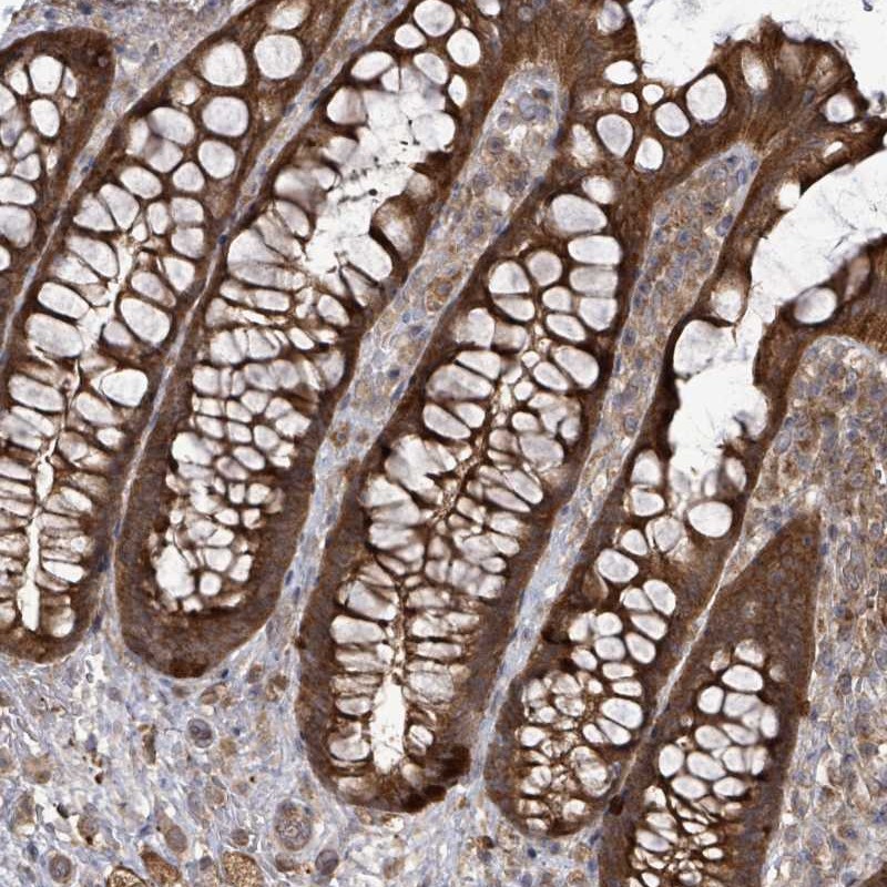
ApplicationsImmunoFluorescence, ImmunoHistoChemistry
Product group Antibodies
ReactivityHuman, Mouse, Rat
TargetEIF3C
Overview
- SupplierReddot Biotech
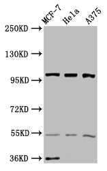
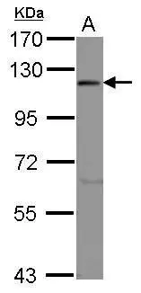
- Product NameEIF3C Polyclonal Antibody
- Delivery Days Customer5
- ApplicationsImmunoFluorescence, ImmunoHistoChemistry
- CertificationResearch Use Only
- Concentration1 mg/ml
- ConjugateUnconjugated
- Gene ID8663
- Target nameEIF3C
- Target descriptioneukaryotic translation initiation factor 3 subunit C
- Target synonymsEIF3CL, EIF3S8, eIF3-p110, eukaryotic translation initiation factor 3 subunit C, cell migration-inducing protein 17, eukaryotic translation initiation factor 3 subunit 8, eukaryotic translation initiation factor 3, subunit 8 (110kD), eukaryotic translation initiation factor 3, subunit 8, 110kDa
- HostRabbit
- IsotypeIgG
- Protein IDQ99613
- Protein NameEukaryotic translation initiation factor 3 subunit C
- Scientific DescriptionComponent of the eukaryotic translation initiation factor 3 (eIF-3) complex, which is required for several steps in the initiation of protein synthesis (PubMed:17581632, PubMed:25849773, PubMed:27462815). The eIF-3 complex associates with the 40S ribosome and facilitates the recruitment of eIF-1, eIF-1A, eIF-2:GTP:methionyl-tRNAi and eIF-5 to form the 43S pre-initiation complex (43S PIC). The eIF-3 complex stimulates mRNA recruitment to the 43S PIC and scanning of the mRNA for AUG recognition. The eIF-3 complex is also required for disassembly and recycling of post-termination ribosomal complexes and subsequently prevents premature joining of the 40S and 60S ribosomal subunits prior to initiation (PubMed:17581632). The eIF-3 complex specifically targets and initiates translation of a subset of mRNAs involved in cell proliferation, including cell cycling, differentiation and apoptosis, and uses different modes of RNA stem-loop binding to exert either translational activation or repression (PubMed:25849773). - This is a EIF3C Polyclonal Antibody from Reddot Biotech. This product is for Research Use Only.
- ReactivityHuman, Mouse, Rat
- Storage Instruction-20°C
- UNSPSC41116161
Related products
Product group Antibodies
Anti-EIF3C AntibodyA31818
ApplicationsImmunoFluorescence, Western Blot, ImmunoHistoChemistry
ReactivityHuman, Mouse, Rat
- SizePrice

Product group Antibodies
Anti-EIF3C Antibody Picoband(r)A06470-1-CARRIER-FREE
ApplicationsFlow Cytometry, Western Blot, ELISA, ImmunoHistoChemistry
ReactivityHuman, Mouse, Rat
TargetEIF3C
- SizePrice
Product group Antibodies
Anti-EIF3C Antibody144-07022
ApplicationsImmunoFluorescence, Western Blot, ImmunoHistoChemistry
ReactivityHuman, Mouse, Rat
TargetEIF3C
- SizePrice
Product group Antibodies

EIF3C / EIF3S8 AntibodyLS-C832147
ApplicationsELISA, ImmunoHistoChemistry
ReactivityHuman, Mouse, Rat
TargetEIF3C
- SizePrice
Product group Antibodies
EIF3C AntibodyCSB-PA857867LA01HU
ApplicationsImmunoFluorescence, Western Blot, ELISA, ImmunoHistoChemistry
ReactivityHuman
TargetEIF3C
- SizePrice
Product group Antibodies
Eif3C Polyclonal AntibodyCAC09690
ApplicationsImmunoFluorescence, Western Blot, ELISA, ImmunoHistoChemistry
TargetEIF3C
- SizePrice
Product group Antibodies
EIF3S8 antibody [C2C3], C-termGTX101801
ApplicationsWestern Blot
ReactivityHuman
TargetEIF3C
- SizePrice
Product group Antibodies
Anti-EIF3C AntibodyHPA050112
ApplicationsWestern Blot, ImmunoHistoChemistry
ReactivityHuman
TargetEIF3C
- SizePrice
Product group Antibodies
Anti-EIF3C AntibodyCAB7022
ApplicationsImmunoFluorescence, Western Blot, ELISA, ImmunoCytoChemistry, ImmunoHistoChemistry, ImmunoHistoChemistry Paraffin
ReactivityHuman
TargetEIF3C
- SizePrice