

ERCC1 / RAD10 (Tumor Progression Marker)(ERCC1/2683), CF594 conjugate, 0.1mg/mL [26628-22-8]
BNC942683
ReactivityBovine, Human, Mouse
Product group Antibodies
TargetERCC1
Overview
- SupplierBiotium
- Product NameERCC1 / RAD10 (Tumor Progression Marker)(ERCC1/2683), CF594 conjugate, 0.1mg/mL [26628-22-8]
- Delivery Days Customer9
- CAS Number26628-22-8
- CertificationResearch Use Only
- ClonalityMonoclonal
- Clone IDERCC1/2683
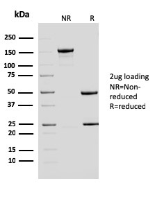
- Concentration0.1 mg/ml
- ConjugateOther Conjugate
- Gene ID2067
- Target nameERCC1
- Target descriptionERCC excision repair 1, endonuclease non-catalytic subunit
- Target synonymsCOFS4, RAD10, UV20, DNA excision repair protein ERCC-1, excision repair cross-complementation group 1, excision repair cross-complementing rodent repair deficiency, complementation group 1 (includes overlapping antisense sequence)
- HostMouse
- IsotypeIgG1
- Protein IDP07992
- Protein NameDNA excision repair protein ERCC-1
- Scientific DescriptionRecognizes a protein of 110 kDa, identified as Excision Repair Cross Complementing 1 (ERCC1). It is a mammalian nucleotide excision repair (NER) enzyme involved in repair of damaged DNA. ERCC1 is a homologous to RAD10 in Saccharomyces cerevisiae, which is required in mitotic intrachromosomal recombination and repair. ERCC1 is required in repair of cisplatin-induced DNA adducts and ultraviolet (UV)-induced DNA damage. High expression of ERCC1 has been linked to tumor progression in a variety of cancers including non-small cell lung cancer (NSCLC), squamous cell carcinoma of the head, ovarian cancer and esophageal cancer. Primary antibodies are available purified, or with a selection of fluorescent CF® Dyes and other labels. CF® Dyes offer exceptional brightness and photostability. Note: Conjugates of blue fluorescent dyes like CF®405S and CF®405M are not recommended for detecting low abundance targets, because blue dyes have lower fluorescence and can give higher non-specific background than other dye colors.
- SourceAnimal
- ReactivityBovine, Human, Mouse
- Storage Instruction2°C to 8°C,RT
- UNSPSC41116161
MSDS
Related products
Product group Antibodies
Anti-ERCC1 AntibodyA285983
ApplicationsImmunoFluorescence, ELISA
ReactivityHuman
- SizePrice
Product group Antibodies
Anti-ERCC1 Antibody144-05291
ApplicationsImmunoFluorescence, Western Blot, ImmunoHistoChemistry
ReactivityHuman, Mouse, Rat
TargetERCC1
- SizePrice

Product group Antibodies
Anti-ERCC1 AntibodyAMAB90871
ApplicationsImmunoCytoChemistry, ImmunoHistoChemistry
ReactivityHuman
TargetERCC1
- SizePrice
Product group Antibodies

ERCC1 Antibody (clone 8K5)LS-C772646
ApplicationsWestern Blot
ReactivityHuman
TargetERCC1
- SizePrice

Product group Antibodies
Anti-ERCC1 Antibody Picoband(r)A00388-2-CARRIER-FREE
ApplicationsWestern Blot, ELISA
ReactivityHuman, Mouse, Rat
TargetERCC1
- SizePrice
Product group Antibodies
ApplicationsFlow Cytometry, Western Blot, ImmunoCytoChemistry
ReactivityHuman, Mouse
TargetERCC1
- SizePrice
Product group Antibodies
ERCC1 Monoclonal AntibodyCSB-MA000236
ApplicationsWestern Blot, ELISA
ReactivityHuman
TargetERCC1
- SizePrice
Product group Antibodies
References

Goat anti-ERCC1EB08190
ApplicationsWestern Blot, ELISA
ReactivityHuman
TargetERCC1
- SizePrice
![ERCC1 antibody [N1C3] detects ERCC1 protein at nucleus by immunofluorescent analysis. Sample: A549 cells were fixed in 4% paraformaldehyde at RT for 15 min. Green: ERCC1 protein stained by ERCC1 antibody [N1C3] (GTX110562) diluted at 1:1000. Blue: Hoechst 33342 staining. Scale bar = 10 μm.](https://www.genetex.com/upload/website/prouct_img/normal/GTX110562/GTX110562_40401_20150324_IFA_w_23060500_388.webp)
Product group Antibodies
ERCC1 antibody [N1C3]GTX110562
ApplicationsImmunoFluorescence, Western Blot, ImmunoCytoChemistry
ReactivityHuman
TargetERCC1
- SizePrice