

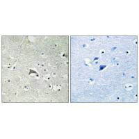
Immunohistochemistry analysis of paraffin-embedded human brain tissue using VEGFR1 antibody.
Immunohistochemistry analysis of paraffin-embedded human brain tissue using VEGFR1 antibody.
FLT1 Antibody
CSB-PA714296
ApplicationsELISA, ImmunoHistoChemistry
Product group Antibodies
ReactivityHuman, Mouse
TargetFLT1
Overview
- SupplierCusabio
- Product NameFLT1 Antibody
- Delivery Days Customer20
- ApplicationsELISA, ImmunoHistoChemistry
- CertificationResearch Use Only
- ClonalityPolyclonal
- ConjugateUnconjugated
- Gene ID2321
- Target nameFLT1
- Target descriptionfms related receptor tyrosine kinase 1
- Target synonymsFLT; FLT-1; fms related tyrosine kinase 1; fms-like tyrosine kinase 1; fms-related tyrosine kinase 1 (vascular endothelial growth factor/vascular permeability factor receptor); tyrosine-protein kinase FRT; tyrosine-protein kinase receptor FLT; vascular endothelial growth factor receptor 1; vascular permeability factor receptor; VEGFR1; VEGFR-1
- HostRabbit
- IsotypeIgG
- Protein IDP17948
- Protein NameVascular endothelial growth factor receptor 1
- Scientific DescriptionTyrosine-protein kinase that acts as a cell-surface receptor for VEGFA, VEGFB and PGF, and plays an essential role in the development of embryonic vasculature, the regulation of angiogenesis, cell survival, cell migration, macrophage function, chemotaxis, and cancer cell invasion. May play an essential role as a negative regulator of embryonic angiogenesis by inhibiting excessive proliferation of endothelial cells. Can promote endothelial cell proliferation, survival and angiogenesis in adulthood. Its function in promoting cell proliferation seems to be cell-type specific. Promotes PGF-mediated proliferation of endothelial cells, proliferation of some types of cancer cells, but does not promote proliferation of normal fibroblasts (in vitro). Has very high affinity for VEGFA and relatively low protein kinase activity; may function as a negative regulator of VEGFA signaling by limiting the amount of free VEGFA and preventing its binding to KDR. Likewise, isoforms lacking a transmembrane domain, such as isoform 2, isoform 3 and isoform 4, may function as decoy receptors for VEGFA. Modulates KDR signaling by forming heterodimers with KDR. Ligand binding leads to the activation of several signaling cascades. Activation of PLCG leads to the production of the cellular signaling molecules diacylglycerol and inositol 1,4,5-trisphosphate and the activation of protein kinase C. Mediates phosphorylation of PIK3R1, the regulatory subunit of phosphatidylinositol 3-kinase, leading to activation of phosphatidylinositol kinase and the downstream signaling pathway. Mediates activation of MAPK1/ERK2, MAPK3/ERK1 and the MAP kinase signaling pathway, as well as of the AKT1 signaling pathway. Phosphorylates SRC and YES1, and may also phosphorylate CBL. Isoform 1 phosphorylates PLCG. Promotes phosphorylation of AKT1 at Ser-473. Promotes phosphorylation of PTK2/FAK1. Isoform 7 has a truncated kinase domain; it increases phosphorylation of SRC at Tyr-418 by unknown means and promotes tumor cell invasion. Shibuya M., Oncogene 5:519-524(1990). Kendall R.L., Proc. Natl. Acad. Sci. U.S.A. 90:10705-10709(1993). The MGC Project Team; Genome Res. 14:2121-2127(2004).
- ReactivityHuman, Mouse
- Storage Instruction-20°C or -80°C
- UNSPSC12352203