

GAD2 / GAD65 (GABAergic Neuronal Marker) (GAD2/1960), CF405S conjugate, 0.1mg/mL [26628-22-8]
BNC041960
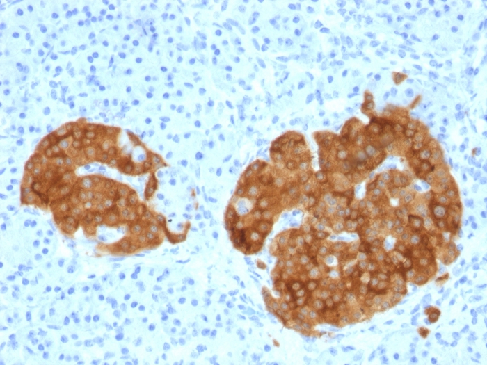
ApplicationsImmunoHistoChemistry, ImmunoHistoChemistry Paraffin
Product group Antibodies
ReactivityBovine, Human, Mouse
TargetGAD2
Overview
- SupplierBiotium
- Product NameGAD2 / GAD65 (GABAergic Neuronal Marker) (GAD2/1960), CF405S conjugate, 0.1mg/mL [26628-22-8]
- Delivery Days Customer9
- ApplicationsImmunoHistoChemistry, ImmunoHistoChemistry Paraffin
- CAS Number26628-22-8
- CertificationResearch Use Only
- ClonalityMonoclonal
- Clone IDGAD2/1960
- Concentration0.1 mg/ml
- ConjugateOther Conjugate
- Gene ID2572
- Target nameGAD2
- Target descriptionglutamate decarboxylase 2
- Target synonymsGAD65, glutamate decarboxylase 2, 65 kDa glutamic acid decarboxylase, GAD-65, Glutamate decarboxylase-2 (pancreas), glutamate decarboxylase 2 (pancreatic islets and brain, 65kDa)
- HostMouse
- IsotypeIgG2b
- Protein IDQ05329
- Protein NameGlutamate decarboxylase 2
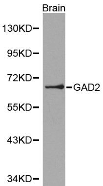
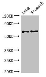
- Scientific DescriptionThis MAb recognizes a protein of 65 kDa, which is identified as glutamic acid decarboxylase 2 (GDA2). It is responsible for catalyzing the production of gamma-aminobutyric acid from L-glutamic acid. There are two forms of glutamic acid decarboxylases (GAD s) that are found in the brain: GAD2 (also known as GAD65) and GAD1 (also known as GAD67). GAD1 and GAD2 are members of the group II decarboxylase family of proteins and are responsible for catalyzing the rate-limiting step in the production of GABA (Z3-aminobutyric acid) from L-glutamic acid. Although both GAD s are found in the brain, GAD2 localizes to synaptic vesicle membranes in nerve terminals, while GAD1 is distributed throughout the cell. A pathogenic role for GAD2 is identified in the human pancreas since it has been identified as an autoantibody and an auto-reactive T cell target in insulin-dependent diabetes. Primary antibodies are available purified, or with a selection of fluorescent CF® Dyes and other labels. CF® Dyes offer exceptional brightness and photostability. Note: Conjugates of blue fluorescent dyes like CF®405S and CF®405M are not recommended for detecting low abundance targets, because blue dyes have lower fluorescence and can give higher non-specific background than other dye colors.
- SourceAnimal
- ReactivityBovine, Human, Mouse
- Storage Instruction2°C to 8°C,RT
- UNSPSC41116161
MSDS
Related products
Product group Antibodies
Anti-GAD2 Antibody144-00971
ApplicationsImmunoFluorescence, Western Blot, ImmunoHistoChemistry
ReactivityHuman, Mouse, Rat
TargetGAD2
- SizePrice

Product group Antibodies
Anti-GAD65/GAD2 Antibody Picoband(r)A03142-1-CARRIER-FREE
ApplicationsWestern Blot, ELISA, ImmunoHistoChemistry
ReactivityHuman, Mouse, Rat
TargetGAD2
- SizePrice

Product group Antibodies
Anti-GAD2 AntibodyAMAB91048
ApplicationsWestern Blot, ImmunoHistoChemistry
ReactivityHuman, Mouse, Rat
TargetGAD2
- SizePrice
Product group Antibodies
Anti-GAD2 AntibodyA28907
ApplicationsWestern Blot, ImmunoHistoChemistry
ReactivityHuman, Mouse, Rat
- SizePrice
Product group Antibodies

Goat anti-GAD2 / GAD65EB06730
ApplicationsWestern Blot, ELISA, ImmunoHistoChemistry
ReactivityCanine, Human, Mouse, Rat
TargetGAD2
- SizePrice
Product group Antibodies
GAD2 AntibodyCSB-PA11159A0RB
ApplicationsWestern Blot, ELISA, ImmunoHistoChemistry
ReactivityHuman, Mouse
TargetGAD2
- SizePrice
Product group Antibodies

GAD65 AntibodyLS-C405593
ApplicationsELISA, ImmunoHistoChemistry
ReactivityHuman
TargetGAD2
- SizePrice
![GAD65 antibody [C2C3], C-term detects GAD65 protein by immunohistochemical analysis. Sample: Frozen sectioned adult mouse retina. Green: GAD65 protein stained by GAD65 antibody [C2C3], C-term (GTX100281) diluted at 1:250. Red: Protein kinase C alpha staining. Blue: Fluoroshield with DAPI (GTX30920).](https://www.genetex.com/upload/website/prouct_img/normal/GTX100281/GTX100281_39456_20160808_IHC-Fr_w_23060100_399.webp)
Product group Antibodies
GAD65 antibody [C2C3], C-termGTX100281
ApplicationsImmunoFluorescence, Western Blot, ImmunoCytoChemistry, ImmunoHistoChemistry, ImmunoHistoChemistry Frozen, ImmunoHistoChemistry Paraffin
ReactivityHuman, Mouse, Rat
TargetGAD2
- SizePrice