

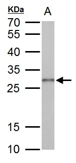
Galectin3 antibody detects Galectin3 protein by western blot analysis. A. 50 μg rat colon lysate/extract 12 % SDS-PAGE Galectin3 antibody (GTX125897) dilution: 1:1000
 and Galectin 3 knockout (KO) HeLa cell extracts (30 μg) were separated by 12% SDS-PAGE, and the membrane was blotted with Galectin 3 antibody (GTX125897) diluted at 1:500. The HRP-conjugated anti-rabbit IgG antibody (GTX213110-01) was used to detect the primary antibody, and the signal was developed with Trident ECL plus-Enhanced.")
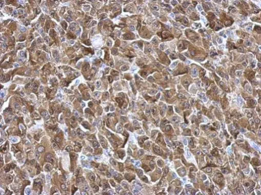
 antibody at 1:200 dilution.
Antigen Retrieval: Trilogy? (EDTA based, pH 8.0) buffer, 15min")
 A: Non-transfected 293T lysate B: LGALS3 transfected 293T lysates 12% SDS PAGE GTX125897 diluted at 1:1000")
 was separated by 12% SDS-PAGE, and the membrane was blotted with Galectin 3 antibody (GTX125897) diluted at 1:1000. The HRP-conjugated anti-rabbit IgG antibody (GTX213110-01) was used to detect the primary antibody.")
 (GTX213110-01) was diluted at 1:10000 and used to detect the primary antibody.")
 A: A549 B: HCT116 12% SDS PAGE GTX125897 diluted at 1:1000")
Galectin3 antibody detects Galectin3 protein by western blot analysis. A. 50 μg rat colon lysate/extract 12 % SDS-PAGE Galectin3 antibody (GTX125897) dilution: 1:1000
Galectin 3 antibody
GTX125897
ApplicationsWestern Blot, ELISA, ImmunoHistoChemistry, ImmunoHistoChemistry Frozen, ImmunoHistoChemistry Paraffin
Product group Antibodies
ReactivityHuman, Mouse, Rat
TargetLGALS3
Overview
- SupplierGeneTex
- Product NameGalectin 3 antibody
- Delivery Days Customer9
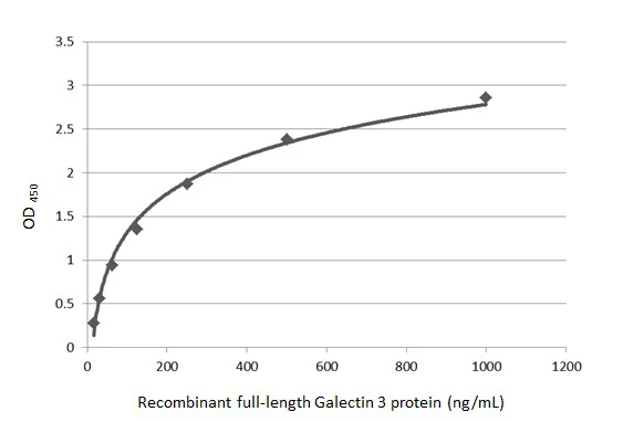
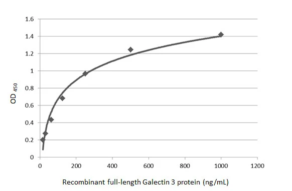
- Application Supplier NoteWB: 1:500-1:3000. IHC-P: 1:100-1:1000. Sandwich ELISA: . *Optimal dilutions/concentrations should be determined by the researcher.Not tested in other applications.
- ApplicationsWestern Blot, ELISA, ImmunoHistoChemistry, ImmunoHistoChemistry Frozen, ImmunoHistoChemistry Paraffin
- CertificationResearch Use Only
- ClonalityPolyclonal
- Concentration1 mg/ml
- ConjugateUnconjugated
- Gene ID3958
- Target nameLGALS3
- Target descriptiongalectin 3
- Target synonymsCBP35, GAL3, GALBP, GALIG, L31, LGALS2, MAC2, galectin-3, 35 kDa lectin, IgE-binding protein, MAC-2 antigen, advanced glycation end-product receptor 3, carbohydrate-binding protein 35, epididymis secretory sperm binding protein, galactose-specific lectin 3, laminin-binding protein, lectin L-29, lectin, galactoside-binding, soluble, 3
- HostRabbit
- IsotypeIgG
- Protein IDP17931
- Protein NameGalectin-3
- Scientific DescriptionThis gene encodes a member of the galectin family of carbohydrate binding proteins. Members of this protein family have an affinity for beta-galactosides. The encoded protein is characterized by an N-terminal proline-rich tandem repeat domain and a single C-terminal carbohydrate recognition domain. This protein can self-associate through the N-terminal domain allowing it to bind to multivalent saccharide ligands. This protein localizes to the extracellular matrix, the cytoplasm and the nucleus. This protein plays a role in numerous cellular functions including apoptosis, innate immunity, cell adhesion and T-cell regulation. Alternate splicing results in multiple transcript variants.
- ReactivityHuman, Mouse, Rat
- Storage Instruction-20°C or -80°C,2°C to 8°C
- UNSPSC12352203
References
- Sbroscia M, Di Gioacchino M, Ascenzi P, et al. Thyroid cancer diagnosis by Raman spectroscopy. Sci Rep. 2020,10(1):13342. doi: 10.1038/s41598-020-70165-0Read this paper
- Li FY, Weng IC, Lin CH, et al. Helicobacter pylori induces intracellular galectin-8 aggregation around damaged lysosomes within gastric epithelial cells in a host O-glycan-dependent manner. Glycobiology. 2019,29(2):151-162. doi: 10.1093/glycob/cwy095Read this paper
- Lin GL, Ting HJ, Tseng TC, et al. Modulation of the mRNA-binding protein HuR as a novel reversal mechanism of epirubicin-triggered multidrug resistance in colorectal cancer cells. PLoS One. 2017,12(10):e0185625. doi: 10.1371/journal.pone.0185625Read this paper
- Brandi J, Dalla Pozza E, Dando I, et al. Secretome protein signature of human pancreatic cancer stem-like cells. J Proteomics. 2016,136:1-12. doi: 10.1016/j.jprot.2016.01.017Read this paper
- Huang HJ, Lin CC, Chou HC, et al. Proteomic analysis of rhein-induced cyt: ER stress mediates cell death in breast cancer cells. Mol Biosyst. 2014,10(12):3086-100. doi: 10.1039/c4mb00451eRead this paper
- Chemaly ER, Kang S, Zhang S, et al. Differential patterns of replacement and reactive fibrosis in pressure and volume overload are related to the propensity for ischaemia and involve resistin. J Physiol. 2013,591(21):5337-55. doi: 10.1113/jphysiol.2013.258731Read this paper
- Chen JY, Chou HC, Chen YH, et al. High glucose-induced proteome alterations in hepatocytes and its possible relevance to diabetic liver disease. J Nutr Biochem. 2013,24(11):1889-910. doi: 10.1016/j.jnutbio.2013.05.006Read this paper
Datasheet
Related products
Product group Antibodies
Anti-Galectin-3 Antibody130-10090
ApplicationsELISA
ReactivityHuman
TargetLGALS3
- SizePrice

Product group Antibodies
Anti-LGALS3 Antibody Picoband(r)A00621-1-CARRIER-FREE
ApplicationsImmunoFluorescence, Western Blot, ELISA, ImmunoCytoChemistry
ReactivityHuman, Mouse, Rat
TargetLGALS3
- SizePrice

Product group Antibodies
ApplicationsImmunoCytoChemistry
ReactivityHuman
TargetLGALS3
- SizePrice
Product group Antibodies
References
Galectin 3 antibodyGTX113486
ApplicationsImmunoFluorescence, Western Blot, ELISA, ImmunoCytoChemistry, ImmunoHistoChemistry, ImmunoHistoChemistry Paraffin
ReactivityCanine, Human, Rat
TargetLGALS3
- SizePrice
![Various whole cell extracts (30 μg) were separated by 12% SDS-PAGE, and the membrane was blotted with Galectin 3 antibody [GT1473] (GTX635151) diluted at 1:1000. The HRP-conjugated anti-mouset IgG antibody (GTX213111-01) was used to detect the primary antibody.](https://www.genetex.com/upload/website/prouct_img/normal/GTX635151/GTX635151_43465_20190308_WB_w_23061202_134.webp)
Product group Antibodies
Galectin 3 antibody [GT1473]GTX635151
ApplicationsWestern Blot, ELISA, ImmunoHistoChemistry, ImmunoHistoChemistry Paraffin
ReactivityHuman
TargetLGALS3
- SizePrice
Product group Antibodies
Galectin 3 antibody [GT759]GTX635152
ApplicationsWestern Blot, ELISA
ReactivityHuman
TargetLGALS3
- SizePrice
Product group Antibodies
Galectin 3 antibody [GT12712]GTX635155
ApplicationsImmunoFluorescence, Western Blot, ELISA, ImmunoCytoChemistry, ImmunoHistoChemistry, ImmunoHistoChemistry Paraffin
ReactivityHuman
TargetLGALS3
- SizePrice
![Various whole cell extracts (30 μg) were separated by 12% SDS-PAGE, and the membrane was blotted with Galectin 3 antibody [GT1188] (GTX635156) diluted at 1:500. The HRP-conjugated anti-mouse IgG antibody (GTX213111-01) was used to detect the primary antibody.](https://www.genetex.com/upload/website/prouct_img/normal/GTX635156/GTX635156_43486_20190222_WB_2_w_23051500_246.webp)
Product group Antibodies
Galectin 3 antibody [GT1188]GTX635156
ApplicationsWestern Blot, ELISA, ImmunoHistoChemistry, ImmunoHistoChemistry Paraffin
ReactivityHuman
TargetLGALS3
- SizePrice
![Wild-type (WT) and Galectin 3 knockout (KO) HeLa cell extracts (30 μg) were separated by 12% SDS-PAGE, and the membrane was blotted with Galectin 3 antibody [HL1878] (GTX637627) diluted at 1:1000. The HRP-conjugated anti-rabbit IgG antibody (GTX213110-01) was used to detect the primary antibody.](https://www.genetex.com/upload/website/prouct_img/normal/GTX637627/GTX637627_T-44830_20221104_WB_KO_watermark_22110919_604.webp)
Product group Antibodies
Galectin 3 antibody [HL1878]GTX637627
ApplicationsImmunoFluorescence, Western Blot, ImmunoCytoChemistry
ReactivityHuman, Mouse
TargetLGALS3
- SizePrice