

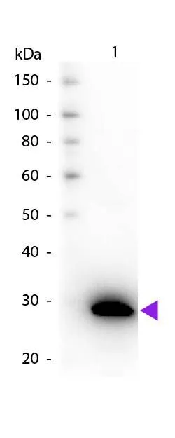
Western blot of Biotin Goat Anti-GFP antibody. Lane 1: GFP. Load: 50 ng per lane. Primary antibody: GFP antibody Biotin conjugated at 1:1,000 for 60 min at RT. Secondary antibody: Peroxidase streptavidin secondary antibody at 1:40,000 for 30 min at RT. Blocking for 30 min at RT. Predicted/Observed size: 28 kDa, 28 kDa for GFP. Other band(s): None.
. Coating : 10 microg/mL")
Western blot of Biotin Goat Anti-GFP antibody. Lane 1: GFP. Load: 50 ng per lane. Primary antibody: GFP antibody Biotin conjugated at 1:1,000 for 60 min at RT. Secondary antibody: Peroxidase streptavidin secondary antibody at 1:40,000 for 30 min at RT. Blocking for 30 min at RT. Predicted/Observed size: 28 kDa, 28 kDa for GFP. Other band(s): None.
GFP antibody (Biotin)
GTX26658
ApplicationsFlow Cytometry, ImmunoFluorescence, ImmunoPrecipitation, Western Blot, ELISA, ImmunoCytoChemistry, ImmunoHistoChemistry, ImmunoHistoChemistry Paraffin, Other Application
Product group Antibodies
ReactivityJellyfish, Other Species
Overview
- SupplierGeneTex
- Product NameGFP antibody (Biotin)
- Delivery Days Customer9
- Application Supplier NoteWB: 1:2000-1:10000. IHC-P: 1:1000-1:5000. ELISA: 1:50000-1:80000. *Optimal dilutions/concentrations should be determined by the researcher.Not tested in other applications.
- ApplicationsFlow Cytometry, ImmunoFluorescence, ImmunoPrecipitation, Western Blot, ELISA, ImmunoCytoChemistry, ImmunoHistoChemistry, ImmunoHistoChemistry Paraffin, Other Application
- CertificationResearch Use Only
- ClonalityPolyclonal
- Concentration1 mg/ml
- ConjugateBiotin
- HostGoat
- IsotypeIgG
- Scientific DescriptionDesigned to detect GFP and its variants in ELISA (sandwich or capture), immunoblotting and immunoprecipitation
- ReactivityJellyfish, Other Species
- Storage Instruction-20°C or -80°C,2°C to 8°C
- UNSPSC12352203
References
- Bettke JA, Tam JW, Montoya V, et al. Inflammatory Monocytes Promote Granuloma-Mediated Control of Persistent Salmonella Infection. Infect Immun. 2022,90(4):e0007022. doi: 10.1128/iai.00070-22Read this paper
- Graef FA, Celiberto LS, Allaire JM, et al. Fasting increases microbiome-based colonization resistance and reduces host inflammatory responses during an enteric bacterial infection. PLoS Pathog. 2021,17(8):e1009719. doi: 10.1371/journal.ppat.1009719Read this paper
- Manko-Prykhoda A, Allain T, Motta JP, et al. Giardia spp. promote the production of antimicrobial peptides and attenuate disease severity induced by attaching and effacing enteropathogens via the induction of the NLRP3 inflammasome. Int J Parasitol. 2020,50(4):263-275. doi: 10.1016/j.ijpara.2019.12.011Read this paper
- Schmitt M, Schewe M, Sacchetti A, et al. Paneth Cells Respond to Inflammation and Contribute to Tissue Regeneration by Acquiring Stem-like Features through SCF/c-Kit Signaling. Cell Rep. 2018,24(9):2312-2328.e7. doi: 10.1016/j.celrep.2018.07.085Read this paper
- Dunlap MD, Howard N, Das S, et al. A novel role for C-C motif chemokine receptor 2 during infection with hypervirulent Mycobacterium tuberculosis. Mucosal Immunol. 2018,11(6):1727-1742. doi: 10.1038/s41385-018-0071-yRead this paper
- Houbracken I, Bouwens L. Acinar cells in the neonatal pancreas grow by self-duplication and not by neogenesis from duct cells. Sci Rep. 2017,7(1):12643. doi: 10.1038/s41598-017-12721-9Read this paper
- Manko A, Motta JP, Cotton JA, et al. Giardia co-infection promotes the secretion of antimicrobial peptides beta-defensin 2 and trefoil factor 3 and attenuates attaching and effacing bacteria-induced intestinal disease. PLoS One. 2017,12(6):e0178647. doi: 10.1371/journal.pone.0178647Read this paper
- Paratore F, Zeidman Kalman T, Rosenfeld T, et al. Isotachophoresis-Based Surface Immunoassay. Anal Chem. 2017,89(14):7373-7381. doi: 10.1021/acs.analchem.7b00725Read this paper
- Morampudi V, Graef FA, Stahl M, et al. Tricellular Tight Junction Protein Tricellulin Is Targeted by the Enteropathogenic Escherichia coli Effector EspG1, Leading to Epithelial Barrier Disruption. Infect Immun. 2017,85(1). doi: 10.1128/IAI.00700-16Read this paper
- Song I, Patel O, Himpe E, et al. Beta Cell Mass Restoration in Alloxan-Diabetic Mice Treated with EGF and Gastrin. PLoS One. 2015,10(10):e0140148. doi: 10.1371/journal.pone.0140148Read this paper