

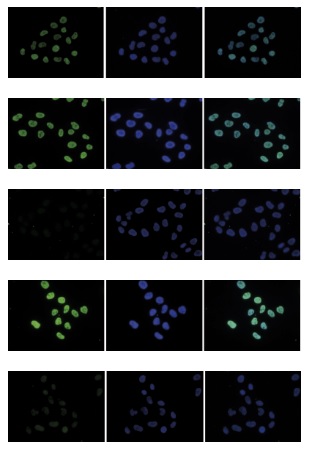
HeLa cells were stained with the Bioss antibody against H4K5,8,12ac (cat. No. bs-53096R) and with DAPI. Cells were fixed with 4% formaldehyde for 10’ and blocked with PBS/TX-100 containing 5% normal goat serum and 1% BSA. Figure A: cells were immunofluorescently labeled with the H4K5,8,12ac antibody (left) diluted 1:500 in blocking solution followed by Alexa488 conjugated secondary antibody. The middle panel shows staining of the nuclei with DAPI. A merge of the two stainings is shown on the right. Figure B, C, D, and E: staining of the cells with the H4K5,8,12ac antibody after incubation of the antibody with 10 ng/μl of the following blocking peptides: H4K5,8,12 unmodified (figure B), H4K5,8,12ac (figure C), H2A.ZK5,7,11ac (figure D) and H4K5,8,12,16ac (figure E).
 and histone extracts (15 μg, lane 2) of HeLa cells, and on 1 μg of recombinant histone H2A, H2B, H3 and H4 (lane 3, 4, 5 and 6, respectively) using the Bioss antibody against H4K5,8,12ac (cat. No. bs-53096R). The antibody was diluted 1:1,000 in TBS-Tween containing 5% skimmed milk.")
, a Dot Blot analysis was performed with peptides containing other histone modifications and the unmodified H4. One hundred to 0.2 pmol of the respective peptides were spotted on a membrane. The antibody was used at a dilution of 1:20,000. Figure shows a high specificity of the antibody for the modification of interest.")
 in antigen-coated wells. The antigen used was a peptide containing the histone modification of interest. By plotting the absorbance against the antibody dilution, the titer of the antibody was estimated to be 1:14,500.")
 on sheared chromatin from 100,000 K562 cells using a ChIP-seq kit. The IP’d DNA was subsequently analyzed on an Illumina Genome Analyzer. Library preparation, cluster generation, and sequencing were performed according to the manufacturer’s instructions. The 36 bp tags were aligned to the human genome using the ELAND algorithm. The figure shows the signal distribution along the complete length of chromosome 2 (figure A) and a zoomin to a 600 kb region (figure B). Figure C and D show the enrichment in two genomic regions on chromosome 3 and 12, respectively, containing EIF4A2 and GAPDH positive controls.")
 and optimized PCR primer sets for qPCR. ChIP was performed with a ChIP- seq kit on sheared chromatin from 100,000 cells. A titration of the antibody consisting of 0.2, 0.5, 1 and 2 μg per ChIP experiment was analyzed. IgG (1 μg/IP) was used as negative IP control. QPCR was performed with primers for the promoter of the active gene EIF4A2 and for a region 1 kb upstream of the GAPDH gene, used as positive controls, and for the inactive MYOD1 gene and the Sat2 satellite repeat region used as negative controls. The figure shows the recovery, expressed as a % of input (the relative amount of immunoprecipitated DNA compared to input DNA after qPCR analysis).")
HeLa cells were stained with the Bioss antibody against H4K5,8,12ac (cat. No. bs-53096R) and with DAPI. Cells were fixed with 4% formaldehyde for 10’ and blocked with PBS/TX-100 containing 5% normal goat serum and 1% BSA. Figure A: cells were immunofluorescently labeled with the H4K5,8,12ac antibody (left) diluted 1:500 in blocking solution followed by Alexa488 conjugated secondary antibody. The middle panel shows staining of the nuclei with DAPI. A merge of the two stainings is shown on the right. Figure B, C, D, and E: staining of the cells with the H4K5,8,12ac antibody after incubation of the antibody with 10 ng/μl of the following blocking peptides: H4K5,8,12 unmodified (figure B), H4K5,8,12ac (figure C), H2A.ZK5,7,11ac (figure D) and H4K5,8,12,16ac (figure E).
H4K5 8 12ac Polyclonal Antibody
BS-53096R
ApplicationsDot Blot, ImmunoFluorescence, ImmunoPrecipitation, Western Blot, ChIP Chromatin ImmunoPrecipitation, ELISA
Product group Antibodies
ReactivityHuman, Mouse
TargetH4C9
Overview
- SupplierBioss Antibodies
- Product NameH4K5 8 12ac Polyclonal Antibody
- Delivery Days Customer16
- ApplicationsDot Blot, ImmunoFluorescence, ImmunoPrecipitation, Western Blot, ChIP Chromatin ImmunoPrecipitation, ELISA
- Applications SupplierWB(1:300-5000), ELISA(1:500-1000), IP(1-2ug), IF(), dot-blot(), ChIP-seq()
- CertificationResearch Use Only
- ClonalityPolyclonal
- Concentration0.75 ug/ul
- ConjugateUnconjugated
- Gene ID8294
- Target nameH4C9
- Target descriptionH4 clustered histone 9
- Target synonymsH4 histone family, member M; H4/m; H4-16; H4C1; H4C11; H4C12; H4C13; H4C14; H4C15; H4C2; H4C3; H4C4; H4C5; H4C6; H4C8; H4FM; H4M; HIST1H4I; histone 1, H4i; Histone 4 family, member M; histone cluster 1 H4 family member i; histone cluster 1, H4i; histone family member; histone H4
- HostRabbit
- Protein IDP62805
- Protein NameHistone H4
- ReactivityHuman, Mouse
- Storage Instruction-20°C
- UNSPSC12352203