

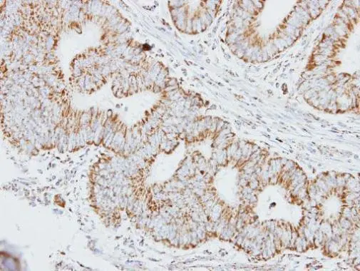
Immunohistochemical analysis of paraffin-embedded human colon carcinoma, using HADHA(GTX113727) antibody at 1:250 dilution.
Antigen Retrieval: Trilogy? (EDTA based, pH 8.0) buffer, 15min
 A: mouse liver 7.5% SDS PAGE GTX113727 diluted at 1:1000")
![HADHA antibody [N2C1], Internal detects HADHA protein at Mitochondia by immunofluorescent analysis. Sample: HepG2 cells were fixed in -20oC 100% MeOH for 5 min. Green: HADHA protein stained by HADHA antibody [N2C1], Internal (GTX113727) diluted at 1:500. Blue: Hoechst 33343 staining.](https://www.genetex.com/upload/website/prouct_img/normal/GTX113727/GTX113727_40142_IFA_w_23060501_967.webp "HADHA antibody [N2C1], Internal detects HADHA protein at Mitochondia by immunofluorescent analysis. Sample: HepG2 cells were fixed in -20oC 100% MeOH for 5 min. Green: HADHA protein stained by HADHA antibody [N2C1], Internal (GTX113727) diluted at 1:500. Blue: Hoechst 33343 staining.")
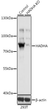
![Non-transfected (–) and transfected (+) 293T whole cell extracts (30 μg) were separated by 7.5% SDS-PAGE, and the membrane was blotted with HADHA antibody [N2C1], Internal (GTX113727) diluted at 1:1000. The HRP-conjugated anti-rabbit IgG antibody (GTX213110-01) was used to detect the primary antibody.](https://www.genetex.com/upload/website/prouct_img/normal/GTX113727/GTX113727_40142_20181026_WB_shRNA_watermark_w_23060501_595.webp "Non-transfected (–) and transfected (+) 293T whole cell extracts (30 μg) were separated by 7.5% SDS-PAGE, and the membrane was blotted with HADHA antibody [N2C1], Internal (GTX113727) diluted at 1:1000. The HRP-conjugated anti-rabbit IgG antibody (GTX213110-01) was used to detect the primary antibody.")
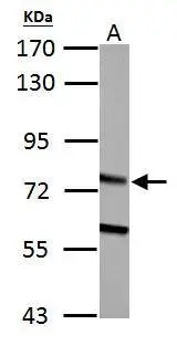
![HADHA antibody [N2C1], Internal detects HADHA protein by Western blot analysis. A.30 μg PC-12 whole cell lysate/extract 7.5 % SDS-PAGE HADHA antibody [N2C1], Internal (GTX113727) dilution: 1:500](https://www.genetex.com/upload/website/prouct_img/normal/GTX113727/GTX113727_40142_WB_R_w_23060501_418.webp "HADHA antibody [N2C1], Internal detects HADHA protein by Western blot analysis. A.30 μg PC-12 whole cell lysate/extract 7.5 % SDS-PAGE HADHA antibody [N2C1], Internal (GTX113727) dilution: 1:500")
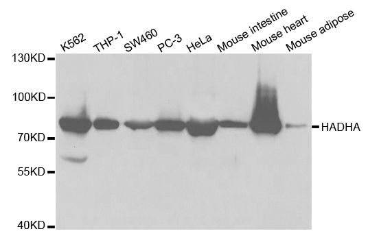
 A: 293T B: A431 (GTX27909) 7.5% SDS PAGE GTX113727 diluted at 1:1000")
![HADHA antibody [N2C1], Internal detects HADHA protein at cytoplasm by immunofluorescent analysis. Sample: HeLa cells were fixed in 4% paraformaldehyde at RT for 15 min. Green: HADHA protein stained by HADHA antibody [N2C1], Internal (GTX113727) diluted at 1:500. Red: alpha Tubulin, a cytoskeleton marker, stained by alpha Tubulin antibody [GT114] (GTX628802) diluted at 1:1000. Blue: Hoechst 33342 staining.](https://www.genetex.com/upload/website/prouct_img/normal/GTX113727/GTX113727_40142_20150410_IFA_w_23060501_386.webp "HADHA antibody [N2C1], Internal detects HADHA protein at cytoplasm by immunofluorescent analysis. Sample: HeLa cells were fixed in 4% paraformaldehyde at RT for 15 min. Green: HADHA protein stained by HADHA antibody [N2C1], Internal (GTX113727) diluted at 1:500. Red: alpha Tubulin, a cytoskeleton marker, stained by alpha Tubulin antibody [GT114] (GTX628802) diluted at 1:1000. Blue: Hoechst 33342 staining.")

Immunohistochemical analysis of paraffin-embedded human colon carcinoma, using HADHA(GTX113727) antibody at 1:250 dilution.
Antigen Retrieval: Trilogy? (EDTA based, pH 8.0) buffer, 15min
HADHA antibody [N2C1], Internal
GTX113727
ApplicationsImmunoFluorescence, Western Blot, ImmunoCytoChemistry, ImmunoHistoChemistry, ImmunoHistoChemistry Paraffin
Product group Antibodies
ReactivityHuman, Mouse, Rat
TargetHADHA
Overview
- SupplierGeneTex
- Product NameHADHA antibody [N2C1], Internal
- Delivery Days Customer9
- Application Supplier NoteWB: 1:500-1:3000. ICC/IF: 1:100-1:1000. IHC-P: 1:100-1:1000. *Optimal dilutions/concentrations should be determined by the researcher.Not tested in other applications.
- ApplicationsImmunoFluorescence, Western Blot, ImmunoCytoChemistry, ImmunoHistoChemistry, ImmunoHistoChemistry Paraffin
- CertificationResearch Use Only
- ClonalityPolyclonal
- Concentration0.37 mg/ml
- ConjugateUnconjugated
- Gene ID3030
- Target nameHADHA
- Target descriptionhydroxyacyl-CoA dehydrogenase trifunctional multienzyme complex subunit alpha
- Target synonymsECHA, GBP, HADH, LCEH, LCHAD, MLCL AT, MTPA, TP-ALPHA, trifunctional enzyme subunit alpha, mitochondrial, 3-ketoacyl-Coenzyme A (CoA) thiolase, alpha subunit, 3-oxoacyl-CoA thiolase, 78 kDa gastrin-binding protein, gastrin-binding protein, hydroxyacyl-CoA dehydrogenase/3-ketoacyl-CoA thiolase/enoyl-CoA hydratase (trifunctional protein), alpha subunit, hydroxyacyl-Coenzyme A dehydrogenase/3-ketoacyl-Coenzyme A thiolase/enoyl-Coenzyme A hydratase (trifunctional protein), alpha subunit, long-chain 2-enoyl-CoA hydratase, long-chain-3-hydroxyacyl-CoA dehydrogenase, mitochondrial long-chain 2-enoyl-Coenzyme A (CoA) hydratase, alpha subunit, mitochondrial long-chain L-3-hydroxyacyl-Coenzyme A (CoA) dehydrogenase, alpha subunit, mitochondrial trifunctional enzyme, alpha subunit, mitochondrial trifunctional protein, alpha subunit, monolysocardiolipin acyltransferase
- HostRabbit
- IsotypeIgG
- Protein IDP40939
- Protein NameTrifunctional enzyme subunit alpha, mitochondrial
- Scientific DescriptionThis gene encodes the alpha subunit of the mitochondrial trifunctional protein, which catalyzes the last three steps of mitochondrial beta-oxidation of long chain fatty acids. The mitochondrial membrane-bound heterocomplex is composed of four alpha and four beta subunits, with the alpha subunit catalyzing the 3-hydroxyacyl-CoA dehydrogenase and enoyl-CoA hydratase activities. Mutations in this gene result in trifunctional protein deficiency or LCHAD deficiency. The genes of the alpha and beta subunits of the mitochondrial trifunctional protein are located adjacent to each other in the human genome in a head-to-head orientation. [provided by RefSeq]
- ReactivityHuman, Mouse, Rat
- Storage Instruction-20°C or -80°C,2°C to 8°C
- UNSPSC41116161
Datasheet
Related products
Product group Antibodies
Anti-HADHA AntibodyA30745
ApplicationsImmunoFluorescence, Western Blot, ImmunoHistoChemistry
ReactivityHuman, Mouse, Rat
- SizePrice

Product group Antibodies
Anti-HADHA Antibody Picoband(r)A03666-2-CARRIER-FREE
ApplicationsFlow Cytometry, ImmunoFluorescence, Western Blot, ELISA, ImmunoCytoChemistry, ImmunoHistoChemistry
ReactivityHuman, Mouse, Rat
TargetHADHA
- SizePrice
Product group Antibodies
Anti-HADHA Antibody144-05346
ApplicationsImmunoFluorescence, Western Blot
ReactivityHuman, Mouse
TargetHADHA
- SizePrice
Product group Antibodies
HADHA Polyclonal AntibodyBS-55092R
ApplicationsImmunoFluorescence, ImmunoPrecipitation, Western Blot, ImmunoCytoChemistry
ReactivityHuman, Mouse, Rat
TargetHADHA
- SizePrice
Product group Antibodies
HADHA AntibodyCSB-PA010118GA01HU
ApplicationsWestern Blot, ELISA, ImmunoHistoChemistry
ReactivityHuman, Mouse, Rat
TargetHADHA
- SizePrice
Product group Antibodies
HADHA antibodyGTX101177
ApplicationsImmunoFluorescence, Western Blot, ImmunoCytoChemistry, ImmunoHistoChemistry, ImmunoHistoChemistry Paraffin
ReactivityHuman, Mouse, Rat
TargetHADHA
- SizePrice
Product group Antibodies

HADHA AntibodyLS-C482426
ApplicationsImmunoFluorescence, Western Blot, ImmunoCytoChemistry, ImmunoHistoChemistry, ImmunoHistoChemistry Paraffin
ReactivityHuman, Mouse, Rat
TargetHADHA
- SizePrice
Product group Antibodies
HADHA antibodyGTX32642
ApplicationsImmunoFluorescence, ImmunoPrecipitation, Western Blot, ImmunoCytoChemistry
ReactivityHuman, Mouse
TargetHADHA
- SizePrice

Product group Antibodies
Anti-HADHA AntibodyHPA015536
ApplicationsWestern Blot, ImmunoCytoChemistry, ImmunoHistoChemistry
ReactivityHuman, Mouse
TargetHADHA
- SizePrice