

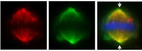
HAUS6 ( IFA using GTX118732 at 1: 500; red) co-localizes at the mitotic spindle with Hice1-GFP (HAUS-GFP; green) in U2OS cells. DAPI staining (blue) shows mitotic chromosomes. Arrows indicate spindle poles.
 A: JurKat 7.5% SDS PAGE GTX118732 diluted at 1:1000 The HRP-conjugated anti-rabbit IgG antibody (GTX213110-01) was used to detect the primary antibody.")
 co-immunoprecipitated with other Augmin components HAUS6 (GTX118732), HAUS2 (GTX118734) and HAUS1 (GTX118733) in U2OS cells. The HRP-conjugated anti-rabbit IgG antibody (GTX213110-01) was used to detect the primary antibody.")
 were separated by 7.5% SDS-PAGE, and the membrane was blotted with HAUS6 antibody (GTX118732) diluted by 1:1000.")
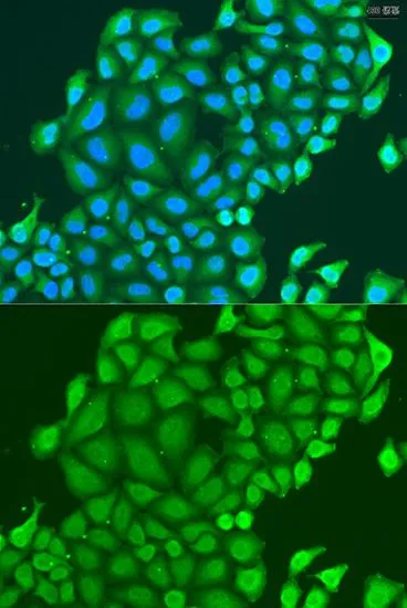
 dilution: 1:500.
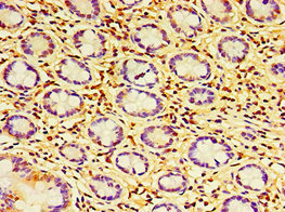
Antigen Retrieval: Trilogy? (EDTA based, pH 8.0) buffer, 15min")


HAUS6 ( IFA using GTX118732 at 1: 500; red) co-localizes at the mitotic spindle with Hice1-GFP (HAUS-GFP; green) in U2OS cells. DAPI staining (blue) shows mitotic chromosomes. Arrows indicate spindle poles.
HAUS6 antibody
GTX118732
ApplicationsImmunoFluorescence, Western Blot, ImmunoCytoChemistry, ImmunoHistoChemistry, ImmunoHistoChemistry Paraffin
Product group Antibodies
ReactivityHuman
TargetHAUS6
Overview
- SupplierGeneTex
- Product NameHAUS6 antibody
- Delivery Days Customer9
- Application Supplier NoteWB: 1:500-1:3000. ICC/IF: 1:100-1:1000. IHC-P: 1:100-1:1000. *Optimal dilutions/concentrations should be determined by the researcher.Not tested in other applications.
- ApplicationsImmunoFluorescence, Western Blot, ImmunoCytoChemistry, ImmunoHistoChemistry, ImmunoHistoChemistry Paraffin
- CertificationResearch Use Only
- ClonalityPolyclonal
- Concentration1 mg/ml
- ConjugateUnconjugated
- Gene ID54801
- Target nameHAUS6
- Target descriptionHAUS augmin like complex subunit 6
- Target synonymsDgt6, FAM29A, HAUS augmin-like complex subunit 6, dim gamma-tubulin homolog, family with sequence similarity 29, member A
- HostRabbit
- IsotypeIgG
- Protein IDQ7Z4H7
- Protein NameHAUS augmin-like complex subunit 6
- Scientific DescriptionHAUS6 is 1 of 8 subunits of the 390-kD human augmin complex, or HAUS complex. The augmin complex was first identified in Drosophila, and its name comes from the Latin verb augmentare, meaning to increase. The augmin complex is a microtubule-binding complex involved in microtubule generation within the mitotic spindle and is vital to mitotic spindle assembly (Goshima et al., 2008 [PubMed 18443220]; Uehara et al., 2009 [PubMed 19369198]).[supplied by OMIM]
- ReactivityHuman
- Storage Instruction-20°C or -80°C,2°C to 8°C
- UNSPSC41116161
Datasheet
Related products
Product group Antibodies
HAUS6 AntibodyCSB-PA767199DSR1HU
ApplicationsELISA, ImmunoHistoChemistry
ReactivityHuman
TargetHAUS6
- SizePrice

Product group Antibodies
Anti-FAM29A/HAUS6 Antibody Picoband(r)A10580-1-CARRIER-FREE
ApplicationsImmunoFluorescence, ImmunoPrecipitation, Western Blot, ELISA, ImmunoCytoChemistry
ReactivityHuman
TargetHAUS6
- SizePrice
Product group Antibodies

HAUS6 / FAM29A AntibodyLS-C831003
ApplicationsELISA, ImmunoHistoChemistry
ReactivityHuman
TargetHAUS6
- SizePrice

Product group Antibodies
Anti-HAUS6 AntibodyHPA020965
ApplicationsImmunoCytoChemistry, ImmunoHistoChemistry
ReactivityHuman
TargetHAUS6
- SizePrice
Product group Antibodies
HAUS6 antibodyGTX65582
ApplicationsImmunoFluorescence, Western Blot, ImmunoCytoChemistry
ReactivityHuman, Mouse
TargetHAUS6
- SizePrice

Product group Antibodies
FAM29A Polyclonal AntibodyBS-9723R
ApplicationsImmunoFluorescence, Western Blot, ImmunoHistoChemistry, ImmunoHistoChemistry Paraffin
ReactivityHuman, Mouse, Rabbit, Rat
- SizePrice
Product group Antibodies
Anti-Mouse HAUS6 Antibody144-04797
ApplicationsImmunoFluorescence, Western Blot
ReactivityHuman, Mouse
TargetHAUS6
- SizePrice