

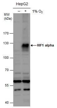
HIF1 alpha antibody detects HIF1 alpha protein by western blot analysis. Un-treated (-) and treated (+, 1% O2 treatment for 24hr) HepG2 whole cell extracts (30 μg) were separated by 7.5% SDS-PAGE, and the membrane was blotted with HIF1 alpha antibody (GTX127309) diluted at 1:1000. The HRP-conjugated anti-rabbit IgG antibody (GTX213110-01) was used to detect the primary antibody.
 dilution: 1:500.")

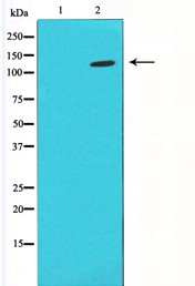
 and treated (+) HeLa whole cell extracts (30 μg) were separated by 7.5% SDS-PAGE, and the membrane was blotted with HIF1 alpha antibody (GTX127309) diluted at 1:1000. The HRP-conjugated anti-rabbit IgG antibody (GTX213110-01) was used to detect the primary antibody.")
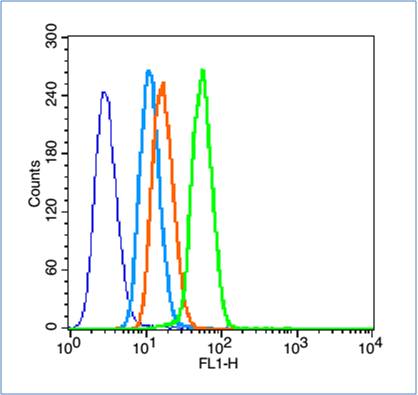
 diluted at 1:200. Blue: Hoechst 33342 staining.")
 and treated (+) HCT116 whole cell extracts (30 μg) were separated by 7.5% SDS-PAGE, and the membrane was blotted with HIF1 alpha antibody (GTX127309) diluted at 1:1000. The HRP-conjugated anti-rabbit IgG antibody (GTX213110-01) was used to detect the primary antibody.")
 and transfected (+) 293T whole cell extracts (30 μg) were separated by 7.5% SDS-PAGE, and the membrane was blotted with HIF1 alpha antibody (GTX127309) diluted at 1:5000. The HRP-conjugated anti-rabbit IgG antibody (GTX213110-01) was used to detect the primary antibody.")
 and treated (+) MCF-7 whole cell extracts (30 μg) were separated by 7.5% SDS-PAGE, and the membrane was blotted with HIF1 alpha antibody (GTX127309) diluted at 1:1000. The HRP-conjugated anti-rabbit IgG antibody (GTX213110-01) was used to detect the primary antibody.")
 and treated (+) HeLa whole cell extracts (30 μg) were separated by 7.5% SDS-PAGE, and the membrane was blotted with HIF1 alpha antibody (GTX127309) diluted at 1:1000. The HRP-conjugated anti-rabbit IgG antibody (GTX213110-01) was used to detect the primary antibody.")
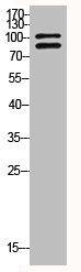
. Western blot analysis was performed using HIF1 alpha antibody (GTX127309). EasyBlot HRP-conjugated anti rabbit IgG antibody (GTX221666-01) was used to detect the primary antibody.")
HIF1 alpha antibody detects HIF1 alpha protein by western blot analysis. Un-treated (-) and treated (+, 1% O2 treatment for 24hr) HepG2 whole cell extracts (30 μg) were separated by 7.5% SDS-PAGE, and the membrane was blotted with HIF1 alpha antibody (GTX127309) diluted at 1:1000. The HRP-conjugated anti-rabbit IgG antibody (GTX213110-01) was used to detect the primary antibody.
HIF1 alpha antibody
GTX127309
ApplicationsImmunoFluorescence, ImmunoPrecipitation, Western Blot, ChIP Chromatin ImmunoPrecipitation, ImmunoCytoChemistry, ImmunoHistoChemistry, ImmunoHistoChemistry Frozen, ImmunoHistoChemistry Paraffin
Product group Antibodies
ReactivityBovine, Human, Mouse, Rabbit, Rat
TargetHIF1A
Overview
- SupplierGeneTex
- Product NameHIF1 alpha antibody
- Delivery Days Customer9
- Application Supplier NoteWB: 1:500-1:3000. ICC/IF: 1:100-1:1000. IHC-P: 1:100-1:1000. IP: 1:100-1:570. *Optimal dilutions/concentrations should be determined by the researcher.Not tested in other applications.
- ApplicationsImmunoFluorescence, ImmunoPrecipitation, Western Blot, ChIP Chromatin ImmunoPrecipitation, ImmunoCytoChemistry, ImmunoHistoChemistry, ImmunoHistoChemistry Frozen, ImmunoHistoChemistry Paraffin
- CertificationResearch Use Only
- ClonalityPolyclonal
- Concentration1.87 mg/ml
- ConjugateUnconjugated
- Gene ID3091
- Target nameHIF1A
- Target descriptionhypoxia inducible factor 1 subunit alpha
- Target synonymsHIF-1-alpha, HIF-1A, HIF-1alpha, HIF1, HIF1-ALPHA, MOP1, PASD8, bHLHe78, hypoxia-inducible factor 1-alpha, ARNT interacting protein, PAS domain-containing protein 8, basic-helix-loop-helix-PAS protein MOP1, class E basic helix-loop-helix protein 78, hypoxia inducible factor 1 alpha subunit, hypoxia inducible factor 1, alpha subunit (basic helix-loop-helix transcription factor), hypoxia-inducible factor1alpha, member of PAS protein 1, member of PAS superfamily 1
- HostRabbit
- IsotypeIgG
- Protein IDQ16665
- Protein NameHypoxia-inducible factor 1-alpha
- Scientific DescriptionHypoxia-inducible factor-1 (HIF1) is a transcription factor found in mammalian cells cultured under reduced oxygen tension that plays an essential role in cellular and systemic homeostatic responses to hypoxia. HIF1 is a heterodimer composed of an alpha subunit and a beta subunit. The beta subunit has been identified as the aryl hydrocarbon receptor nuclear translocator (ARNT). This gene encodes the alpha subunit of HIF-1. Overexpression of a natural antisense transcript (aHIF) of this gene has been shown to be associated with nonpapillary renal carcinomas. Two alternative transcripts encoding different isoforms have been identified. [provided by RefSeq]
- ReactivityBovine, Human, Mouse, Rabbit, Rat
- Storage Instruction-20°C or -80°C,2°C to 8°C
- UNSPSC41116161
Datasheet
Related products
Product group Antibodies
Anti-HIF1A AntibodyA37506
ApplicationsWestern Blot, ImmunoHistoChemistry
ReactivityHuman, Mouse, Rat
- SizePrice
Product group Antibodies
anti-Hif-1 alpha (human), mAb (ANC10G3)ANC-335-020
ApplicationsFlow Cytometry, ELISA
ReactivityHuman
TargetHIF1A
- SizePrice
Product group Antibodies

HIF1A / HIF1 Alpha AntibodyLS-C831817
ApplicationsELISA, ImmunoHistoChemistry
ReactivityHuman, Mouse
TargetHIF1A
- SizePrice

Product group Antibodies
Anti-HIF-1 alpha/HIF1A Antibody Picoband(r)A00013-1-CARRIER-FREE
ApplicationsWestern Blot, ELISA
ReactivityHuman
TargetHIF1A
- SizePrice
Product group Antibodies
References
HIF-1 Alpha Polyclonal AntibodyBS-0737R
ApplicationsFlow Cytometry, ImmunoFluorescence, ImmunoPrecipitation, Western Blot, ELISA, ImmunoCytoChemistry, ImmunoHistoChemistry, ImmunoHistoChemistry Frozen, ImmunoHistoChemistry Paraffin
ReactivityChicken, Human, Mouse, Rat
TargetHIF1A
- SizePrice
Product group Antibodies
HIF1A AntibodyCSB-PA002906
ApplicationsWestern Blot, ELISA, ImmunoHistoChemistry
ReactivityHuman, Mouse, Rat
TargetHIF1A
- SizePrice
Product group Antibodies
Hif1A Polyclonal AntibodyCAC07003
ApplicationsImmunoFluorescence, ELISA, ImmunoHistoChemistry
TargetHIF1A
- SizePrice
![IHC-P analysis of human glioblastoma multiforme tissue using GTX30105 HIF1 alpha antibody [H1alpha67].](https://www.genetex.com/upload/website/prouct_img/normal/GTX30105/GTX30105_532_IHC-P_w_23060722_139.webp)
Product group Antibodies
References
HIF1 alpha antibody [H1alpha67]GTX30105
ApplicationsGel Shift Assay, Flow Cytometry, ImmunoFluorescence, ImmunoPrecipitation, Western Blot, ChIP Chromatin ImmunoPrecipitation, ELISA, ImmunoCytoChemistry, ImmunoHistoChemistry, ImmunoHistoChemistry Frozen, ImmunoHistoChemistry Paraffin, Other Application
ReactivityBovine, Ferret, Human, Mammals, Monkey, Mouse, Porcine, Primate, Rabbit, Rat, Sheep, Xenopus, Yeast
TargetHIF1A
- SizePrice
![IHC-P analysis of human kidney tissue using GTX30115 HIF1 alpha antibody [H1alpha67].](https://www.genetex.com/upload/website/prouct_img/normal/GTX30115/GTX30115_310_IHC-P_w_23060722_205.webp)
Product group Antibodies
References
HIF1 alpha antibody [H1alpha67]GTX30115
ApplicationsGel Shift Assay, Flow Cytometry, ImmunoFluorescence, ImmunoPrecipitation, Western Blot, ChIP Chromatin ImmunoPrecipitation, ELISA, ImmunoCytoChemistry, ImmunoHistoChemistry, ImmunoHistoChemistry Frozen, ImmunoHistoChemistry Paraffin
ReactivityAvian, Bovine, Canine, Ferret, Human, Monkey, Mouse, Porcine, Primate, Rat, Sheep
TargetHIF1A
- SizePrice