

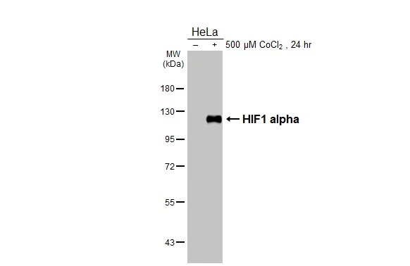
Untreated (–) and treated (+) HeLa whole cell extract (30 μg) were separated by 7.5% SDS-PAGE, and the membrane was blotted with HIF1 alpha antibody [HL3154] (GTX640664) diluted at 1:1000. The HRP-conjugated anti-rabbit IgG antibody (GTX213110-01) was used to detect the primary antibody.
![Untreated (–) and treated (+) NIH-3T3 whole cell extract (30 μg) were separated by 7.5% SDS-PAGE, and the membrane was blotted with HIF1 alpha antibody [HL3154] (GTX640664) diluted at 1:1000. The HRP-conjugated anti-rabbit IgG antibody (GTX213110-01) was used to detect the primary antibody.](https://www.genetex.com/upload/website/prouct_img/normal/GTX640664/GTX640664_T-45481_20240726_WB_M_treatment_CoCl2_24080622_544.webp "Untreated (–) and treated (+) NIH-3T3 whole cell extract (30 μg) were separated by 7.5% SDS-PAGE, and the membrane was blotted with HIF1 alpha antibody [HL3154] (GTX640664) diluted at 1:1000. The HRP-conjugated anti-rabbit IgG antibody (GTX213110-01) was used to detect the primary antibody.")
![Untreated (–) and treated (+) Rat2 whole cell extracts (30 μg) were separated by 7.5% SDS-PAGE, and the membrane was blotted with HIF1 alpha antibody [HL3154] (GTX640664) diluted at 1:1000. The HRP-conjugated anti-rabbit IgG antibody (GTX213110-01) was used to detect the primary antibody.](https://www.genetex.com/upload/website/prouct_img/normal/GTX640664/GTX640664_T-45481_20240809_WB_R_treatment_CoCl2_24081300_722.webp "Untreated (–) and treated (+) Rat2 whole cell extracts (30 μg) were separated by 7.5% SDS-PAGE, and the membrane was blotted with HIF1 alpha antibody [HL3154] (GTX640664) diluted at 1:1000. The HRP-conjugated anti-rabbit IgG antibody (GTX213110-01) was used to detect the primary antibody.")
![HIF1 alpha antibody [HL3154] detects HIF1 alpha protein by immunofluorescent analysis. Sample: Mock and treated HeLa cells were fixed in ice-cold MeOH for 5 min. Green: HIF1 alpha stained by HIF1 alpha antibody [HL3154] (GTX640664) diluted at 1:500. Red: alpha Tubulin, a cytoskeleton marker, stained by alpha Tubulin antibody [GT114] (GTX628802) diluted at 1:1000.](https://www.genetex.com/upload/website/prouct_img/normal/GTX640664/GTX640664_T-45481_20240816_ICC_IF_treatment_CoCl2_24082301_938.webp "HIF1 alpha antibody [HL3154] detects HIF1 alpha protein by immunofluorescent analysis. Sample: Mock and treated HeLa cells were fixed in ice-cold MeOH for 5 min. Green: HIF1 alpha stained by HIF1 alpha antibody [HL3154] (GTX640664) diluted at 1:500. Red: alpha Tubulin, a cytoskeleton marker, stained by alpha Tubulin antibody [GT114] (GTX628802) diluted at 1:1000.")
![Untreated (–) and treated (+) HeLa whole cell extracts (30 μg) were separated by 7.5% SDS-PAGE, and the membranes were blotted with HIF1 alpha antibody [HL3154] (GTX640664) diluted at 1:1000 and competitor's antibody (Competitor) diluted at 1:1000. The HRP-conjugated anti-rabbit IgG antibody (GTX213110-01) was used to detect the primary antibody. *The competitor is not affiliated with GeneTex and does not endorse this product.](https://www.genetex.com/upload/website/prouct_img/normal/GTX640664/GTX640664_45537_20240927_WB_treatment_CoCl2_competitor_watermark_24100318_634.webp "Untreated (–) and treated (+) HeLa whole cell extracts (30 μg) were separated by 7.5% SDS-PAGE, and the membranes were blotted with HIF1 alpha antibody [HL3154] (GTX640664) diluted at 1:1000 and competitor's antibody (Competitor) diluted at 1:1000. The HRP-conjugated anti-rabbit IgG antibody (GTX213110-01) was used to detect the primary antibody. *The competitor is not affiliated with GeneTex and does not endorse this product.")
![HIF1 alpha antibody [HL3154] detects HIF1 alpha protein by immunohistochemical analysis. Sample: Paraffin-embedded mouse tissues. HIF1 alpha stained by HIF1 alpha antibody [HL3154] (GTX640664) diluted at 1:100. Antigen Retrieval: Citrate buffer, pH 6.0, 15 min Corresponding RNA levels (RPKM) in the tissues are based on NCBI database.](https://www.genetex.com/upload/website/prouct_img/normal/GTX640664/GTX640664_45537_20250206_IHC-P_M_Multiple_RPKM_25022521_779.webp "HIF1 alpha antibody [HL3154] detects HIF1 alpha protein by immunohistochemical analysis. Sample: Paraffin-embedded mouse tissues. HIF1 alpha stained by HIF1 alpha antibody [HL3154] (GTX640664) diluted at 1:100. Antigen Retrieval: Citrate buffer, pH 6.0, 15 min Corresponding RNA levels (RPKM) in the tissues are based on NCBI database.")
![HIF1 alpha antibody [HL3154] detects HIF1 alpha protein by immunohistochemical analysis. Sample: Paraffin-embedded rat tissues. HIF1 alpha stained by HIF1 alpha antibody [HL3154] (GTX640664) diluted at 1:100. Antigen Retrieval: Citrate buffer, pH 6.0, 15 min](https://www.genetex.com/upload/website/prouct_img/normal/GTX640664/GTX640664_45537_20250314_IHC-P_M_Multiple_RPKM_25032719_991.webp "HIF1 alpha antibody [HL3154] detects HIF1 alpha protein by immunohistochemical analysis. Sample: Paraffin-embedded rat tissues. HIF1 alpha stained by HIF1 alpha antibody [HL3154] (GTX640664) diluted at 1:100. Antigen Retrieval: Citrate buffer, pH 6.0, 15 min")
![MDCK whole cell extract (30 μg) was separated by 5% SDS-PAGE, and the membrane was blotted with HIF1 alpha antibody [HL3154] (GTX640664) diluted at 1:1000. The HRP-conjugated anti-rabbit IgG antibody (GTX213110-01) was used to detect the primary antibody.](https://www.genetex.com/upload/website/prouct_img/normal/GTX640664/GTX640664_45537_20250321_WB_D_25032719_786.webp "MDCK whole cell extract (30 μg) was separated by 5% SDS-PAGE, and the membrane was blotted with HIF1 alpha antibody [HL3154] (GTX640664) diluted at 1:1000. The HRP-conjugated anti-rabbit IgG antibody (GTX213110-01) was used to detect the primary antibody.")
Untreated (–) and treated (+) HeLa whole cell extract (30 μg) were separated by 7.5% SDS-PAGE, and the membrane was blotted with HIF1 alpha antibody [HL3154] (GTX640664) diluted at 1:1000. The HRP-conjugated anti-rabbit IgG antibody (GTX213110-01) was used to detect the primary antibody.
HIF1 alpha antibody [HL3154]
GTX640664
ApplicationsImmunoFluorescence, Western Blot, ImmunoCytoChemistry, ImmunoHistoChemistry, ImmunoHistoChemistry Paraffin
Product group Antibodies
ReactivityHuman, Mouse, Rat
TargetHIF1A
Overview
- SupplierGeneTex
- Product NameHIF1 alpha antibody [HL3154]
- Delivery Days Customer7
- Application Supplier NoteWB: 1:500-1:3000. *Optimal dilutions/concentrations should be determined by the researcher.Not tested in other applications.
- ApplicationsImmunoFluorescence, Western Blot, ImmunoCytoChemistry, ImmunoHistoChemistry, ImmunoHistoChemistry Paraffin
- CertificationResearch Use Only
- ClonalityMonoclonal
- Clone IDHL3154
- Concentration1 mg/ml
- ConjugateUnconjugated
- Gene ID3091
- Target nameHIF1A
- Target descriptionhypoxia inducible factor 1 subunit alpha
- Target synonymsHIF-1-alpha, HIF-1A, HIF-1alpha, HIF1, HIF1-ALPHA, MOP1, PASD8, bHLHe78, hypoxia-inducible factor 1-alpha, ARNT interacting protein, PAS domain-containing protein 8, basic-helix-loop-helix-PAS protein MOP1, class E basic helix-loop-helix protein 78, hypoxia inducible factor 1 alpha subunit, hypoxia inducible factor 1, alpha subunit (basic helix-loop-helix transcription factor), hypoxia-inducible factor1alpha, member of PAS protein 1, member of PAS superfamily 1
- HostRabbit
- IsotypeIgG
- Protein IDQ16665
- Protein NameHypoxia-inducible factor 1-alpha
- Scientific DescriptionThis gene encodes the alpha subunit of transcription factor hypoxia-inducible factor-1 (HIF-1), which is a heterodimer composed of an alpha and a beta subunit. HIF-1 functions as a master regulator of cellular and systemic homeostatic response to hypoxia by activating transcription of many genes, including those involved in energy metabolism, angiogenesis, apoptosis, and other genes whose protein products increase oxygen delivery or facilitate metabolic adaptation to hypoxia. HIF-1 thus plays an essential role in embryonic vascularization, tumor angiogenesis and pathophysiology of ischemic disease. Alternatively spliced transcript variants encoding different isoforms have been identified for this gene. [provided by RefSeq, Jul 2011]
- ReactivityHuman, Mouse, Rat
- Storage Instruction-20°C or -80°C,2°C to 8°C
- UNSPSC41116161
Datasheet
Related products
Product group Antibodies
HIF1A AntibodyCSB-PA002906
ApplicationsWestern Blot, ELISA, ImmunoHistoChemistry
ReactivityHuman, Mouse, Rat
TargetHIF1A
- SizePrice

Product group Antibodies
Anti-HIF-1 alpha/HIF1A Antibody Picoband(r)A00013-1-CARRIER-FREE
ApplicationsWestern Blot, ELISA
ReactivityHuman
TargetHIF1A
- SizePrice
Product group Antibodies
Anti-HIF1A AntibodyA37506
ApplicationsWestern Blot, ImmunoHistoChemistry
ReactivityHuman, Mouse, Rat
- SizePrice
Product group Antibodies

HIF1A / HIF1 Alpha AntibodyLS-C831817
ApplicationsELISA, ImmunoHistoChemistry
ReactivityHuman, Mouse
TargetHIF1A
- SizePrice

Product group Antibodies
Anti-HIF1A AntibodyHPA000907
ApplicationsImmunoCytoChemistry
ReactivityHuman
TargetHIF1A
- SizePrice
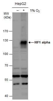
![Untreated (–) and treated (+) HCT116 whole cell extracts (30 μg) were separated by 5% SDS-PAGE, and the membrane was blotted with HIF1 alpha antibody [HL3011] (GTX640424) diluted at 1:1000. The HRP-conjugated anti-rabbit IgG antibody (GTX213110-01) was used to detect the primary antibody.](https://www.genetex.com/upload/website/prouct_img/normal/GTX640424/GTX640424_T-45425_20240614_WB_treatment_hypoxia_24061802_276.webp)
Product group Antibodies
HIF1 alpha antibody [HL3011]GTX640424
ApplicationsImmunoFluorescence, Western Blot, ImmunoCytoChemistry, ImmunoHistoChemistry, ImmunoHistoChemistry Paraffin
ReactivityHuman, Mouse
TargetHIF1A
- SizePrice
Product group Antibodies
HIF1 alpha antibodyGTX127309
ApplicationsImmunoFluorescence, ImmunoPrecipitation, Western Blot, ChIP Chromatin ImmunoPrecipitation, ImmunoCytoChemistry, ImmunoHistoChemistry, ImmunoHistoChemistry Frozen, ImmunoHistoChemistry Paraffin
ReactivityBovine, Human, Mouse, Rabbit, Rat
TargetHIF1A
- SizePrice
![HIF1 alpha antibody [GT10211] detects HIF1 alpha protein at nucleus by immunofluorescent analysis. Sample: Mock and treated HeLa cells were fixed in 4% paraformaldehyde at RT for 15 min. Green: HIF1 alpha stained by HIF1 alpha antibody [GT10211] (GTX628480) diluted at 1:500. Blue: Fluoroshield with DAPI (GTX30920).](https://www.genetex.com/upload/website/prouct_img/normal/GTX628480/GTX628480_44685_20220916_ICC_IF_treatment_CoCl2_22110201_404.webp)
Product group Antibodies
HIF1 alpha antibody [GT10211]GTX628480
ApplicationsImmunoFluorescence, ImmunoPrecipitation, Western Blot, ImmunoCytoChemistry, ImmunoHistoChemistry, ImmunoHistoChemistry Paraffin
ReactivityHuman, Rat
TargetHIF1A
- SizePrice
![Untreated (–) and treated (+) NIH-3T3 whole cell extracts (30 μg) were separated by 5% SDS-PAGE, and the membrane was blotted with HIF1 alpha antibody [GT122] (GTX629766) diluted at 1:1000. The HRP-conjugated anti-mouse IgG antibody (GTX213111-01) was used to detect the primary antibody, and the signal was developed with Trident ECL plus-Enhanced.](https://www.genetex.com/upload/website/prouct_img/normal/GTX629766/GTX629766_44615_20220318_WB_M_treatment_CoCl2_w_23051500_576.webp)
Product group Antibodies
HIF1 alpha antibody [GT122]GTX629766
ApplicationsImmunoFluorescence, Western Blot, ImmunoCytoChemistry
ReactivityHuman, Mouse
TargetHIF1A
- SizePrice