

IRF2BP1 Antibody (aa109-122)
LS-C139734
ApplicationsWestern Blot, ELISA
Product group Antibodies
TargetIRF2BP1
Overview
- SupplierLifeSpan BioSciences
- Product NameIRF2BP1 Antibody (aa109-122)
- Delivery Days Customer23
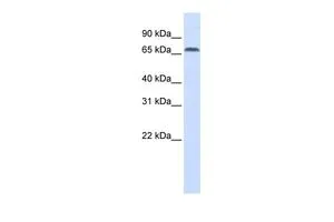
- Application Supplier NotePeptide ELISA: antibody detection limit dilution 1:32000. Western blot: Approx 75kD band observed in lysates of cell line HepG2 (calculated MW of 61.7kD according to NP_056464.1). Recommended concentration: 0.1-0.3 ug/ml.. Peptide-ELISA (1:32000), WB (0.1 - 0.3 µg/ml) Peptide ELISA: antibody detection limit dilution 1:32000. Western blot: Approx 75kD band observed in lysates of cell line HepG2 (calculated MW of 61.7kD according to NP_056464.1). Recommended concentration: 0.1-0.3 ug/ml.
- ApplicationsWestern Blot, ELISA
- CertificationResearch Use Only
- ClonalityPolyclonal
- Concentration0.5 mg/ml
- ConjugateUnconjugated
- Estimated Purity...
- Gene ID26145
- Target nameIRF2BP1
- Target descriptioninterferon regulatory factor 2 binding protein 1
- Target synonymsinterferon regulatory factor 2-binding protein 1; IRF-2-binding protein 1; probable E3 ubiquitin-protein ligase IRF2BP1; probable RING-type E3 ubiquitin transferase IRF2BP1
- HostGoat
- Storage Instruction-20°C
- UNSPSC12352203
Related products
Product group Antibodies
ApplicationsImmunoPrecipitation, Western Blot
TargetIRF2BP1
- SizePrice
Product group Antibodies
IRF2BP1 antibody, InternalGTX45301
ApplicationsWestern Blot
TargetIRF2BP1
- SizePrice
Product group Antibodies
IRF2BP1 Polyclonal AntibodyCAC13403
ApplicationsImmunoFluorescence, ELISA, ImmunoHistoChemistry
TargetIRF2BP1
- SizePrice
Product group Antibodies
IRF2BP1 AntibodyCSB-PA815541LA01HU
ApplicationsImmunoFluorescence, ELISA, ImmunoHistoChemistry
ReactivityHuman
TargetIRF2BP1
- SizePrice

Product group Antibodies
Anti-IRF2BP1 AntibodyHPA042164
ApplicationsWestern Blot, ImmunoHistoChemistry
ReactivityHuman
TargetIRF2BP1
- SizePrice

Product group Antibodies
Anti-IRF2BP1 Antibody Picoband(r)A11406-2-CARRIER-FREE
ApplicationsFlow Cytometry, ImmunoFluorescence, ImmunoPrecipitation, Western Blot, ELISA, ImmunoCytoChemistry, ImmunoHistoChemistry
TargetIRF2BP1
- SizePrice
Product group Antibodies

ApplicationsWestern Blot, ELISA
TargetIRF2BP1
- SizePrice
Product group Antibodies

IRF2BP1 AntibodyLS-C830181
ApplicationsELISA, ImmunoHistoChemistry
TargetIRF2BP1
- SizePrice