

JNK1/2/3 Polyclonal Antibody
RD71379A
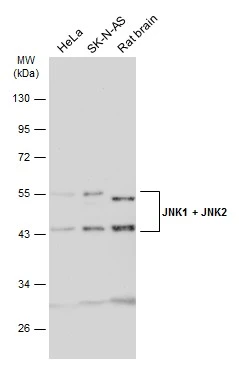
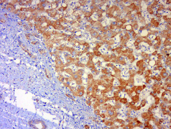
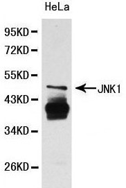
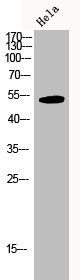
ApplicationsImmunoFluorescence, Western Blot, ELISA, ImmunoHistoChemistry, ImmunoHistoChemistry Paraffin
Product group Antibodies
ReactivityChicken, Human, Mouse, Rat
TargetMAPK8
Overview
- SupplierReddot Biotech
- Product NameJNK1/2/3 Polyclonal Antibody
- Delivery Days Customer5
- ApplicationsImmunoFluorescence, Western Blot, ELISA, ImmunoHistoChemistry, ImmunoHistoChemistry Paraffin
- CertificationResearch Use Only
- ClonalityPolyclonal
- Concentration1 mg/ml
- ConjugateUnconjugated
- Gene ID5599
- Target nameMAPK8
- Target descriptionmitogen-activated protein kinase 8
- Target synonymsJNK, JNK-46, JNK1, JNK1A2, JNK21B1/2, PRKM8, SAPK1, SAPK1c, mitogen-activated protein kinase 8, JUN N-terminal kinase, MAP kinase 8, c-Jun N-terminal kinase 1, stress-activated protein kinase 1, stress-activated protein kinase 1c
- HostRabbit
- IsotypeIgG
- Scientific Descriptionc-Jun N-terminal kinases (JNKs) phosphorylate and augment transcriptional activity of c-Jun. JNKs originate from three genes that yield ten isoforms through alternative mRNA splicing, including JNK1Oe+/-1,JNK1beta1, JNK2Oe+/-1, JNK2beta1 and JNK3Oe+/-1, which represent the p46 isoforms, and JNK1Oe+/-2, JNK1beta2, JNK2Oe+/-2, JNK2beta2 and JNK3beta2, which represent the p54 isoforms. JNKs coordinate cell responses to stress and influence regulation of cell growth and ransformation. The human JNK1 (PRKM8, SAPK1, MAPK8) gene maps to chromosome 10q11.22 and shares 83% amino acid identity with JNK2. JNK1 is necessary for normal activation and differentiation of CD4 helper T (TH) cells into TH1 and TH2 effector cells. Capsaicin activates JNK1 and p38 in Ras-transformed human breast epithelial cells. Nitrogen oxides (NOx) upregulate JNK1 in addition to c-Fos, c-Jun and other signaling kinases, including MEKK1 and p38.
- ReactivityChicken, Human, Mouse, Rat
- Storage Instruction-20°C
- UNSPSC12352203
Related products
Product group Antibodies
Anti-MAPK8 Antibody144-00288
ApplicationsImmunoFluorescence, Western Blot, ImmunoHistoChemistry
ReactivityHuman, Mouse
TargetMAPK8
- SizePrice
Product group Antibodies
References
JNK1 + JNK2 antibodyGTX133806
ApplicationsWestern Blot, ImmunoHistoChemistry, ImmunoHistoChemistry Paraffin
ReactivityHuman, Rat
TargetMAPK8
- SizePrice
Product group Antibodies
MAPK8 Polyclonal AntibodyCAC14610
ApplicationsImmunoFluorescence, Western Blot, ELISA, ImmunoHistoChemistry
TargetMAPK8
- SizePrice
Product group Antibodies
References
JNK1 + JNK3 Polyclonal AntibodyBS-0501R
ApplicationsFlow Cytometry, ImmunoFluorescence, Western Blot, ELISA, ImmunoCytoChemistry, ImmunoHistoChemistry, ImmunoHistoChemistry Frozen, ImmunoHistoChemistry Paraffin
ReactivityBovine, Canine, Chicken, Human, Mouse, Porcine, Rabbit, Rat
TargetMAPK8
- SizePrice
Product group Antibodies
Anti-JNK1 AntibodyA29715
ApplicationsWestern Blot
ReactivityHuman, Mouse, Rat
- SizePrice

Product group Antibodies
References
ApplicationsImmunoFluorescence, ImmunoPrecipitation, Western Blot, ImmunoCytoChemistry
ReactivityHuman, Mouse, Rat
TargetMAPK8
- SizePrice
Product group Antibodies
MAPK8/MAPK9/MAPK10 AntibodyCSB-PA003084
ApplicationsImmunoFluorescence, Western Blot, ELISA, ImmunoHistoChemistry
ReactivityHuman, Mouse, Rat
TargetMAPK8
- SizePrice
Product group Antibodies

ApplicationsELISA
ReactivityHuman
TargetMAPK8
- SizePrice