

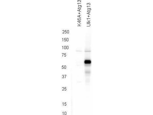
Western blot using Rockland's affinity purified anti-ATG13 pS318 antibody shows detection of phosphorylated ATG13 in 293T cells engineered to coexpress Ulk1 and Atg13 (Ulk1 + Atg13). In the left lane was loaded kinase-dead hypophosphorylated Ulk1-K46A mutant + ATG13. The right lane contains the 293T Ulk1 + ATG13 lysate and shows detection at approximately 57 kDa. The antibody was purified and resolved by SDS-PAGE, then transferred to nitrocellulose membrane. The membrane was blocked with 5% Blotto (p/n B501-0500) and probed with the primary antibody at 1µg/mL overnight at 4°C. After washing, the membrane was probed with Goat Anti-Rabbit HRP secondary 1:5000 in detection buffer (p/n MB-070) for 45 minutes at room temperature. In collaboration with Charles Dorsey at Eli Lilly, Indianapolis, IN and John Cleveland at Scripps, Jupiter, FL.
Western blot using Rockland's affinity purified anti-ATG13 pS318 antibody shows detection of phosphorylated ATG13 in 293T cells engineered to coexpress Ulk1 and Atg13 (Ulk1 + Atg13). In the left lane was loaded kinase-dead hypophosphorylated Ulk1-K46A mutant + ATG13. The right lane contains the 293T Ulk1 + ATG13 lysate and shows detection at approximately 57 kDa. The antibody was purified and resolved by SDS-PAGE, then transferred to nitrocellulose membrane. The membrane was blocked with 5% Blotto (p/n B501-0500) and probed with the primary antibody at 1µg/mL overnight at 4°C. After washing, the membrane was probed with Goat Anti-Rabbit HRP secondary 1:5000 in detection buffer (p/n MB-070) for 45 minutes at room temperature. In collaboration with Charles Dorsey at Eli Lilly, Indianapolis, IN and John Cleveland at Scripps, Jupiter, FL.
KIAA0652 (ATG13) Rabbit Polyclonal Antibody
TA397435S
ApplicationsFlow Cytometry, Western Blot, ELISA
Product group Antibodies
ReactivityHuman
TargetATG13
Overview
- SupplierOriGene
- Product NameKIAA0652 (ATG13) Rabbit Polyclonal Antibody
- Delivery Days Customer14
- ApplicationsFlow Cytometry, Western Blot, ELISA
- CertificationResearch Use Only
- ClonalityPolyclonal
- Gene ID9776
- Target nameATG13
- Target descriptionautophagy related 13
- Target synonymsKIAA0652, PARATARG8, autophagy-related protein 13, ATG13 autophagy related 13 homolog
- HostRabbit
- Protein IDO75143
- Protein NameAutophagy-related protein 13
- Scientific DescriptionATG13 phospho S318 Antibody
- ReactivityHuman
- Storage Instruction-20°C
- UNSPSC12352203
MSDS
Related products
Product group Antibodies
ATG13 AntibodyCSB-PA012203ESR1HU
ApplicationsELISA, ImmunoHistoChemistry
ReactivityHuman
TargetATG13
- SizePrice
Product group Antibodies
Anti-ATG13 AntibodyA28634
ApplicationsImmunoFluorescence, Western Blot, ImmunoHistoChemistry
ReactivityHuman, Mouse, Rat
- SizePrice
Product group Antibodies

KIAA0652 / ATG13 AntibodyLS-C331070
ApplicationsImmunoFluorescence, Western Blot, ImmunoHistoChemistry
ReactivityHuman, Mouse, Rat
TargetATG13
- SizePrice
Product group Antibodies
ATG13 Polyclonal AntibodyCAC14853
ApplicationsWestern Blot, ELISA, ImmunoHistoChemistry
TargetATG13
- SizePrice

Product group Antibodies
Anti-KIAA0652/ATG13 Antibody Picoband(r)PB9480-CARRIER-FREE
ApplicationsWestern Blot
ReactivityHamster, Human, Mouse
TargetATG13
- SizePrice
Product group Antibodies
ATG13 antibodyGTX65842
ApplicationsImmunoFluorescence, Western Blot, ImmunoCytoChemistry, ImmunoHistoChemistry, ImmunoHistoChemistry Paraffin
ReactivityHuman, Rat
TargetATG13
- SizePrice
Product group Antibodies
Anti-ATG13 Antibody144-00690
ApplicationsImmunoFluorescence, Western Blot, ImmunoHistoChemistry
ReactivityHuman, Mouse, Rat
TargetATG13
- SizePrice
Product group Antibodies
ATG13 Polyclonal AntibodyBS-22897R
ApplicationsImmunoFluorescence, ImmunoHistoChemistry, ImmunoHistoChemistry Frozen, ImmunoHistoChemistry Paraffin
ReactivityCanine, Chicken, Human, Mouse, Porcine, Rabbit, Rat, Sheep
TargetATG13
- SizePrice