

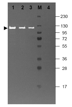
Figure 1. Western blotting using Fluorescein conjugated anti-Beta-galactosidase antibody shows a band at ~117 kDa (lanes 1 - 3) corresponding to 60 ng, 30 ng and 15 ng, respectively of b-Gal present in partially purified preparations (arrowhead). Lane 4 shows no cross reactivity with proteins present in a non-specific control E. coli lysate. Proteins were resolved on a 4-20% Tris-glycine gel by SDS-PAGE and transferred to Nitrocellulose and blocking using blocking buffer for fluorescent Western blotting. The membrane was probed with Fluorescein conjugated anti-Beta-galactosidase (. F) diluted to 1/10,000. Reaction occurred for 2 hours at room temperature. Molecular weight estimation was made by comparison to a prestained MW marker in lane M. Fluorescence image was captured using the VersaDoc® Imaging System. Other detection systems will yield similar results.
Figure 1. Western blotting using Fluorescein conjugated anti-Beta-galactosidase antibody shows a band at ~117 kDa (lanes 1 - 3) corresponding to 60 ng, 30 ng and 15 ng, respectively of b-Gal present in partially purified preparations (arrowhead). Lane 4 shows no cross reactivity with proteins present in a non-specific control E. coli lysate. Proteins were resolved on a 4-20% Tris-glycine gel by SDS-PAGE and transferred to Nitrocellulose and blocking using blocking buffer for fluorescent Western blotting. The membrane was probed with Fluorescein conjugated anti-Beta-galactosidase (. F) diluted to 1/10,000. Reaction occurred for 2 hours at room temperature. Molecular weight estimation was made by comparison to a prestained MW marker in lane M. Fluorescence image was captured using the VersaDoc® Imaging System. Other detection systems will yield similar results.
lacZ Rabbit Polyclonal Antibody

R1064
ApplicationsWestern Blot, ELISA, ImmunoHistoChemistry
Product group Antibodies
ReactivityBacteria
TargetlacZ
Overview
- SupplierOriGene
- Product NamelacZ Rabbit Polyclonal Antibody
- Delivery Days Customer14
- ApplicationsWestern Blot, ELISA, ImmunoHistoChemistry
- CertificationResearch Use Only
- ClonalityPolyclonal
- Gene ID945006
- Target namelacZ
- Target descriptionbeta-galactosidase
- Target synonymsb0344; beta-galactosidase; ECK0341
- HostRabbit
- Scientific DescriptionlacZ rabbit polyclonal antibody, Serum
- ReactivityBacteria
- UNSPSC12352203