

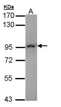
Sample (30 μg of whole cell lysate) A: Molt-4 (GTX27912) 7.5% SDS PAGE GTX109519 diluted at 1:10000 The HRP-conjugated anti-rabbit IgG antibody (GTX213110-01) was used to detect the primary antibody.
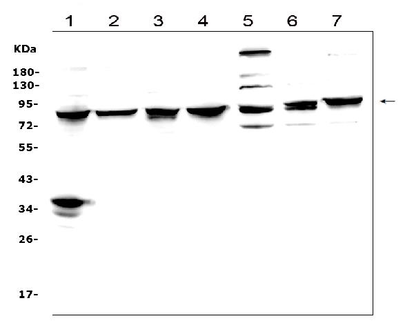
 were separated by 7.5% SDS-PAGE, and the membrane was blotted with MAD1 antibody (GTX109519) diluted by 1:5000.")
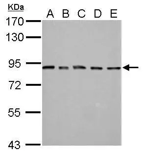
 dilution: 1:3000 The HRP-conjugated anti-rabbit IgG antibody (GTX213110-01) was used to detect the primary antibody.")
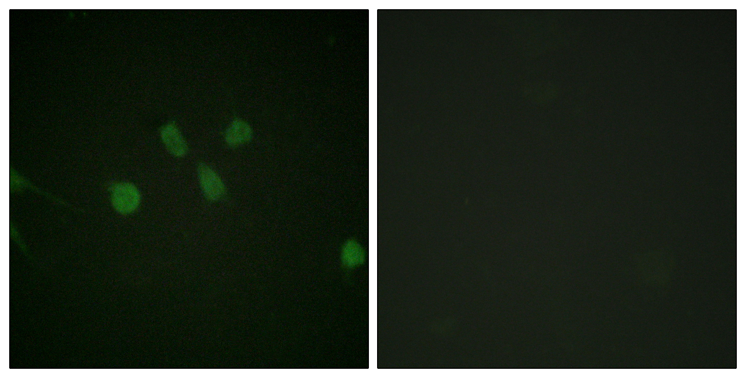
![MAD1 antibody detects MAD1 protein at cytoplasm and nucleus by immunofluorescent analysis. Sample: HeLa cells were fixed in 4% paraformaldehyde at RT for 15 min. Green: MAD1 protein stained by MAD1 antibody (GTX109519) diluted at 1:500. Red: HEC1, a kinetochore marker, stained by HEC1 antibody [9G3.23] (GTX70268) diluted at 1:500. Blue: Hoechst 33342 staining.](https://www.genetex.com/upload/website/prouct_img/normal/GTX109519/GTX109519_40030_20150410_IFA_w_23060500_496.webp "MAD1 antibody detects MAD1 protein at cytoplasm and nucleus by immunofluorescent analysis. Sample: HeLa cells were fixed in 4% paraformaldehyde at RT for 15 min. Green: MAD1 protein stained by MAD1 antibody (GTX109519) diluted at 1:500. Red: HEC1, a kinetochore marker, stained by HEC1 antibody [9G3.23] (GTX70268) diluted at 1:500. Blue: Hoechst 33342 staining.")
. Western blot analysis was performed using MAD1 antibody (GTX109519). EasyBlot anti-Rabbit IgG (GTX221666-01) was used as a secondary reagent.")
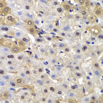
 antibody at 1:100 dilution.
Antigen Retrieval: Trilogy? (EDTA based, pH 8.0) buffer, 15min")
 dilution: 1:3000 The HRP-conjugated anti-rabbit IgG antibody (GTX213110-01) was used to detect the primary antibody.")


Sample (30 μg of whole cell lysate) A: Molt-4 (GTX27912) 7.5% SDS PAGE GTX109519 diluted at 1:10000 The HRP-conjugated anti-rabbit IgG antibody (GTX213110-01) was used to detect the primary antibody.
MAD1 antibody
GTX109519
ApplicationsImmunoFluorescence, ImmunoPrecipitation, Western Blot, ChIP Chromatin ImmunoPrecipitation, ImmunoCytoChemistry, ImmunoHistoChemistry, ImmunoHistoChemistry Paraffin
Product group Antibodies
ReactivityHuman, Mammals, Mouse, Rat
TargetMAD1L1
Overview
- SupplierGeneTex
- Product NameMAD1 antibody
- Delivery Days Customer9
- Application Supplier NoteWB: 1:500-1:20000. ICC/IF: 1:100-1:1000. IHC-P: 1:100-1:1000. IP: 1:100-1:500. *Optimal dilutions/concentrations should be determined by the researcher.Not tested in other applications.
- ApplicationsImmunoFluorescence, ImmunoPrecipitation, Western Blot, ChIP Chromatin ImmunoPrecipitation, ImmunoCytoChemistry, ImmunoHistoChemistry, ImmunoHistoChemistry Paraffin
- CertificationResearch Use Only
- ClonalityPolyclonal
- Concentration1 mg/ml
- ConjugateUnconjugated
- Gene ID8379
- Target nameMAD1L1
- Target descriptionmitotic arrest deficient 1 like 1
- Target synonymsMAD1, MVA7, PIG9, TP53I9, TXBP181, mitotic spindle assembly checkpoint protein MAD1, MAD1 mitotic arrest deficient like 1, MAD1-like protein 1, mitotic arrest deficient 1-like protein 1, mitotic checkpoint MAD1 protein homolog, mitotic-arrest deficient 1, yeast, homolog-like 1, tax-binding protein 181, tumor protein p53 inducible protein 9
- HostRabbit
- IsotypeIgG
- Protein IDQ9Y6D9
- Protein NameMitotic spindle assembly checkpoint protein MAD1
- Scientific DescriptionMAD1L1 is a component of the mitotic spindle-assembly checkpoint that prevents the onset of anaphase until all chromosome are properly aligned at the metaphase plate. MAD1L1 functions as a homodimer and interacts with MAD2L1. MAD1L1 may play a role in cell cycle control and tumor suppression. Three transcript variants encoding the same protein have been found for this gene. [provided by RefSeq]
- ReactivityHuman, Mammals, Mouse, Rat
- Storage Instruction-20°C or -80°C,2°C to 8°C
- UNSPSC41116161
Datasheet
Related products
Product group Antibodies
Phospho-MAD1L1 (S428) AntibodyCSB-PA060282
ApplicationsELISA, ImmunoHistoChemistry
ReactivityHuman
TargetMAD1L1
- SizePrice
Product group Antibodies
Anti-MAD1 AntibodyA101385
ApplicationsImmunoFluorescence, ELISA
ReactivityHuman
- SizePrice

Product group Antibodies
Anti-MAD1L1 AntibodyHPA003635
ApplicationsWestern Blot, ImmunoHistoChemistry
ReactivityHuman
TargetMAD1L1
- SizePrice
Product group Antibodies

MAD1L1 / MAD1 AntibodyLS-C400863
ApplicationsWestern Blot, ELISA
ReactivityHuman, Mouse
TargetMAD1L1
- SizePrice
Product group Antibodies
Anti-MAD1/MAD1L1 Antibody Picoband(r)PB9262-CARRIER-FREE
ApplicationsFlow Cytometry, ImmunoFluorescence, Western Blot, ImmunoCytoChemistry, ImmunoHistoChemistry
ReactivityHuman, Mouse, Rat
TargetMAD1L1
- SizePrice
Product group Antibodies
MAD1 antibodyGTX105079
ApplicationsImmunoFluorescence, ImmunoPrecipitation, Western Blot, ImmunoCytoChemistry, ImmunoHistoChemistry, ImmunoHistoChemistry Paraffin
ReactivityHuman
TargetMAD1L1
- SizePrice
Product group Antibodies
MAD1 antibody [9B10]GTX48641
ApplicationsImmunoPrecipitation, Western Blot, ELISA
ReactivityHuman
TargetMAD1L1
- SizePrice
Product group Antibodies
MAD1 antibodyGTX32710
ApplicationsImmunoFluorescence, Western Blot, ImmunoCytoChemistry, ImmunoHistoChemistry, ImmunoHistoChemistry Paraffin
ReactivityHuman, Mouse
TargetMAD1L1
- SizePrice
Product group Antibodies
MAD1 Recombinant AntibodyBSM-62140R
ApplicationsWestern Blot
ReactivityHuman, Mouse, Rat
TargetMAD1L1
- SizePrice
Product group Antibodies
Anti-MAD1L1 Antibody144-01153
ApplicationsImmunoFluorescence, Western Blot, ImmunoHistoChemistry
ReactivityHuman, Mouse
TargetMAD1L1
- SizePrice